Research Article - (2014) Volume 3, Issue 1
Scientific disciplines normally thought of as outside the sphere of medical science have experienced advances over the past twenty years that currently have profound implications for medical care and medical research. Evolutionary and developmental biology, complexity and chaos science, along with the Human Genome Project and spinoff projects, have dramatically altered our understanding of how the human body functions and what can be expected from research methods like animal modeling. In this article, I summarize these advances and examine one historical medical development, the Blalock-Taussig shunt, in order to ascertain whether historically accepted representations of this development are consistent with current knowledge. I also examine an ongoing technology-intra-arterial stent development-to compare human data to the known animal data and place this comparison in the context of the aforementioned advances.
Keywords: Animal models; Biological complex systems; Blalock-Taussig; Predictive value; Stents; Trans-Species Modeling Theory
The use of animals as models for humans in biomedical research is ethically controversial [1-6]. Less controversial is the scientific merit of using such models. I believe this general lack of concern for the scientific validity of animal models is the result of an inattention to exactly how the models are being used in contradistinction to what is being claimed by the animal model community. Table 1 lists nine categories of animal use in science and research. In this article, I will explore some theoretical concerns of animal models from the fields of complex systems and evolutionary biology, and present empirical evidence that support the theoretical concerns. I will then present a frequently cited historical surgical advance attributed to animal models, and a current invasive procedure also attributed to animal models in order to examine the utility of animal models in these advances.
| 1. | Animals are used as predictive models of humans for research into such diseases as cancer and AIDS. |
| 2. | Animals are used as predictive models of humans for testing drugs or other chemicals. |
| 3. | Animals are used as “spare parts”, such as when a person receives an aortic valve from a pig. |
| 4. | Animals are used as bioreactors or factories, such as for the production of insulin or monoclonal antibodies, or to maintain the supply of a virus. |
| 5. | Animals and animal tissues are used to study basic physiological principles. |
| 6. | Animals are used in education to educate and train medical students and to teach basic principles of anatomy in high school biology classes. |
| 7. | Animals are used as a modality for ideas or as a heuristic device, which is a component of basic science research. |
| 8. | Animals are used in research designed to benefit other animals of the same species or breed. |
| 9. | Animals are used in research in order to gain knowledge for knowledge sake. |
Table 1: Nine categories of animal use in science and research [7].
In preparing this manuscript, I have been cognizant of, or tried to adhere to, the following:
1. One of the main problems in assessing historical advances in science is the lack of primary sources. While we have that problem even in evaluating relatively recent medical discoveries, another more relevant problem is lack of primary or secondary sources that critically evaluate older developments or discoveries. Most sources offer merely a narrative that has been accepted by a group associated with the advance. Rarely does “yet another review” of the development of X yield any really new information.
2. By critical evaluation, I mean using the fundamentals of critical thinking [7-11] and specifically evaluating the premises and claims of the discoverer or developer regarding how the discovery happened. Frequently, the medical researcher responsible for the advance engages in fallacious reasoning in the form of post hoc ergo propter hoc-after this, therefore because of this. This fallacy, along with others, is frequently encountered when authors describe the history of a discovery. There are some good explanations for why this occurs. Rarely does a researcher present the real life events of the discovery [12]. Furthermore, all discoveries take place in the context of societal norms and the ideology of the investigator. These things need to be taken into account when assessing what was necessary for the discovery, what was not necessary, and what was actually misleading but the scientist persevered despite the misleading data. The distinction between necessary and sufficient is rarely addressed in assessing past advances in biomedical science. My concern for these first two items is consistent with skepticism and critical thinking, as well as philosophy of science [13-16]. Evaluating the past with this in mind also allows for Bayesian analysis [17].
3. In light of the above, I also attempt to put past discoveries in the context of what is possible or probable in current science. In other words, if a scientist discovers something new about the universe and credits this discovery to his use of homeopathic remedies or n-rays we can discount those claims as current science disallows any therapeutic effect of homeopathy [18,19] and n-rays and psychic powers do not exist. Likewise, if a past discovery was credited to animal models and current science, which is more advanced than the science at the time of the discovery, is unable to use animal models in this fashion, then we can conclude that there were probably other, more important factors leading to this discovery. It may also be possible to conclude that the discovery simply followed from the laws of physics.
I acknowledge that the above assertion offers more in terms of determining what did not assist in a discovery or development than in isolating what exactly did happen, when it happened, or why it was the most important factor. Such analyses are valuable but so are analyses that exclude specific factors. I will outline other relevant factors that must be considered when assessing biomedical advances in the introductory paragraphs that follow.
Medical science has advanced by numerous and varied methods [20]. Hard work, expertise in the area, dedication, and pursuing an idea that should be right, based on other scientific principles, sometimes in the face of dogged criticism, have figured heavily in medical advances. But other factors and circumstances have also been important. These include accidents [21], as was the case for the discovery of penicillin, the discovery of radioactivity, and the discovery that one could inject dye directly into the coronary arteries.
As Pasteur acknowledged, chance, similar to accidents, favors only the prepared mind. A scientist notices something unexpected, realizes the importance of it, and pursues it to its logical conclusion. Barbara McClintock’s jumping genes are an example of this type of scientific advance. Reasoning by analogy is frequently applied in science. For example, because Krebs had previously discovered a biochemical process that was a cycle-the ornithine cycle for urea synthesis-he reasoned by analogy that perhaps the biochemical process of respiration (now known as the Krebs cycle) would also involve a continuous loop. Discoveries can also happen because technologies that were previously unavailable become accessible. Watson and Crick had access to Franklin’s x-ray crystallography images and hence were able to deduce the structure of DNA.
Historically, many surgical advances have occurred because irrational biases were shown to be wrong. (Current medical practice also suffers from irrational biases [22].) For example, the notions that one could not operate on basal ganglia [23-25] or the heart persisted for decades. Theodor Billroth was quoted (possibly apocryphally) as saying that “any surgeon who wishes to preserve the respect of his colleagues would never attempt to suture the heart” [26]. Regardless of the validity of the quote, it represents the sentiment of the era. Rehn first sutured the heart of a gardener dying from a puncture [27,28] in the late 1800s, thereby ushering in the era of cardiac surgery. Robert Gross was the first to tie off the ductus arteriosus, the vessel connecting the pulmonary circulation and the aorta in fetuses. See Figure 1. The ductus remaining open long after birth is harmful to the baby. Gross had learned much about the anatomy of this condition when he was a first-year resident in pathology and performed autopsies on children who had died from the condition [26]. Gross ligated a patent ductus arteriosus on August 26, 1938. Like all arguments based on the assertion “we have always done it this way,” Gross was warned that ligating the duct would result in catastrophe. A junior surgeon at the time, he had been explicitly told not to perform this new and dangerous operation by his chief. When the chief went out of town however, Gross ignored the admonition and performed the operation anyway. Upon returning home and learning of Gross’s operation, the chief fired him but hired him back at a later date.
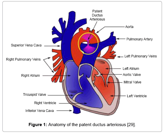
Figure 1: Anatomy of the patent ductus arteriosus [29].
Others surgical advances were the result of accidents, necessity (usually on the battlefield), or were the rational outgrowths of knowledge available at the time. The blood vessels severed in amputations were not historically ligated or cauterized as they are today. During the American Civil War, surgeons decided the stop the bleeding, in part because they were literally up their ankles in blood and it was interfering with further operations. (Paré had actually pioneered this in the 17th century.) In 1953, Cooper discovered that the area of the brain supplied by the anterior choroidal artery was involved in tremors when he accidently severed the artery [29-31]. Advances in technology led to the use of fiberoptics and the development of laparoscopic surgeries.
An example of an operation that was inspired in one case by human observation and in another case by animal studies was the correction of the coarctation of the aorta (CoA). Clarence Crafoord of Sweden successfully operated on a child with a CoA on October 19, 1944. Crafoord claimed that he got the idea for how to repair the CoA when operating on another patient for a similar problem. The abovementioned Gross, who performed the operation a few weeks after Crafoord but whose article was published first in the scientific literature, accused Crafoord of stealing his idea for how to repair the CoA, an idea that had apparently come been developed in the dog lab. Crafoord’s originality has, however, been vindicated by others [26]. Animal studies were thus not necessary for this advance.
Some operations seem to obey the laws of physics in terms of fluid dynamics but, in reality, fail to provide benefit for the patient. For example, in the mid-1960s extracranial-intracranial (EC-IC) bypass procedures for inoperable carotid artery disease were tested and perfected on dogs and rabbits [32,33]. The operation was projected to channel blood from outside the brain to inside the brain, hence the name. Neurosurgeons performed a large number of the procedures ostensibly in order to prevent strokes that otherwise would have resulted from compromised blood supply to the brain. Since the blood vessels in the head but outside the brain rarely exhibit atherosclerosis, they should be ideal candidates for increasing the supply of blood to the brain. EC-IC was practiced for 20 years before anyone questioned it. In 1985, Barnett and Peerless reported on a large study that revealed the procedure actually did more harm than good [34]. More patients died or suffered strokes because of the operation than were helped as a result of it. Further studies confirmed this [34-37].
Other operations have been performed on humans because they were effective in animals. Based largely on studies in dogs, the internal mammary artery (IMA) was ligated in hopes of increasing blood flow to the heart, the thinking being that collateral circulation would form from the ligated IMA and the coronaries [38-44]. Versions of the IMA ligation were performed and some surgeons reported good outcomes [43,45]. Even Reader’s Digest proclaimed the procedure a success [46]. However, more thorough studies did not replicate the promising animal data. The procedures were abandoned when a double-blind study was performed with half the patients undergoing ligation of the IMA and the others receiving a skin incision without IMA ligation. There was no difference in outcome [45]. Interestingly, further studies revealed that the IMA ligation did not protect dogs when the anterior descending coronary artery was ligated [47]. This reveals yet another problem when attempting to interpret older studies-there may have been methodological problems that were unappreciated at the time.
In 1935, Beck placed a section of the trapezius muscle onto the heart in the area of a blocked coronary artery. In dogs this procedure appeared to provide a new blood supply [48]. Based on the dog studies, Beck performed the operation on seven patients with limited success. He also pioneered other operations on the human heart based on dog models, but these were equally unsuccessful [49-53]. Vineberg then modified the muscle procedure, based again on studies in dogs, by making a tunnel into the area supplied by the blocked artery and connecting the internal mammary artery to the tunnel [54] Based on the supposed success of the Vineberg procedure, other cardiac surgeons attempted to modify the operation, but again with limited success [55]. Eventually, there was angiographic evidence that the Vineberg procedure provided some arterial flow to the coronaries, but this did not fully vindicate the procedure as surgeons had conflicting outcomes [56]. Ultimately it was replaced by direct IMA to coronary grafting [57]. (Although, there has recently been renewed interest in the Vineberg procedure [58]).
Serendipity has been important in surgical advances. Charles Bailey pioneered the first mitral valve commissurotomy based on his experience selling women’s girdles in his school days and his observation of a deer’s heart beating for hours after it was removed from the body [26]. Bailey also practiced extensively on 60 dogs. The concept was sound, however the knowledge gained as a result of this experience can be questioned as four of his first five patients died during or shortly after the operation [59]. These examples raise the question: What objective criteria can be applied in order to ascertain the effectiveness of a practice? The anecdotes are interesting in their own right, but also because, unlike much of history, we have the words of some of the surgeons involved. However, it is plain that many of them, if not all, did not attempt to break down which facet of their research, or the research of others, was the most important aspect of the process, which elements were unnecessary, and which were vital. For the most part, they simply recounted the developmental process, saying: “We did X followed by Y along with Z, and after that we did A and B and C,” and so on. This type of narrative predisposes one to use the post hoc ergo propter hoc fallacy. There is no real critical thinking surrounding the recitation of the advance. “What did we learn from X? Did Y translate from animals to humans? Could the discovery have been achieved via a different route?”
The people who risked the lives of their patients, along with their own reputations and careers, and saw patients die with regularity were not philosophers of science. They were reflective only in terms of: “Why did this patient die and what can we do differently next time?” Such thinking is typical of surgical advances, as well as medical advances in general. But the application of real critical thinking in terms of the advance is usually lacking. Moreover, after the advance is made, curiosity seems abated. For example, there are many unanswered questions about poliovirus, such as why were the outbreaks seasonal? These questions were simply no longer of interest after the vaccines were developed.
In order to fully appreciate and critique discoveries, critical thinking and philosophy of science skills are required. Thinking critically raises several questions when considering the role of animals as models for humans in surgical procedures. Did the use of animals merely allow surgeons to discard previously held irrational bias? Could the irrational belief have been discarded on another basis? Which portion of the operation, if any, was dependent upon animal models? Was the development of the operation based on knowledge currently available? Did performing the operation on animals result in unwarranted confidence in the procedure?
Where I to suffer a severe, traumatic, cardiac insult, I would want a surgeon who was talented at cardiac surgery, not a philosopher of science. However, were I to want an analysis of past discoveries, I would want the skills one learns in philosophy of science. It is not disparaging to say that the people who made the discoveries or invented the machines might not have had the best grasp of the philosophy of science. Many surgeons credited their experience in the cadaver lab with a breakthrough while other credited their experience operating on dogs. Others just thought it all made sense and proceeded to do what they did. The entire early history of cardiac surgery, from the 1930s through the 1950s, in terms of exactly which research technique or aspect of clinical acumen led to which breakthrough is hazy to say the least. Further, many surgeons expressed the opinion that the dog lab gave them confidence to proceed while also emphasizing that they were in entirely new territory and that each surgical decision in humans had unknown consequences [26]. This is not what one would expect from people who thought the dog lab was exactly like the operating room. Nevertheless, research with animals, specifically dogs, was commonplace.
In evaluating the role of animal models, one must also consider the history of operations that, while successful in animals, did work out well for humans. For example, Elliot Cutler performed the first successful operation to relieve mitral stenosis, but the procedure was initially shown to have an 86% mortality rate (six out of seven patients died) despite excellent success in the lab [26,60,61]. More examples could be given [26,62-66]. Almost all of the surgeons who performed new operations on the heart had a very high mortality rate for that procedure before it was more or less perfected. I do not think this fact is, in any way, indicative of unethical behavior or callousness on the part of the surgeons. New procedures, especially on organs such as the heart, are going to have a high mortality rate, and I see nothing that can be done even today, that was not also done back then, to change that fact. These high mortality rates do call into question the value of the dog model, however.
I surveyed the relevant scientific literature regarding the BT shunt, intra-arterial stents, complex systems, evolutionary biology including evolutionary and developmental biology (evo devo), genetics and genomics, personalized medicine, the history of cardiovascular surgery, the history of interventional cardiology, empirical data relating to animal models of human disease and drug response, and the philosophy of science. I did this in order to formulate a theoretical framework in which to critically examine the historical development of the BT shunt and the ongoing development of intra-arterial stents. I then sought to derive conclusions regarding animal models in general as well as the development of these specific invasive cardiovascular interventions.
Predictions in science
A frequent claim regarding animal models is that they have predictive value for human response to perturbations such as disease and drugs. Some even claim that basic research that employs animals is predictive for humans, although such a claim is the antithesis of basic research [67]. Andrew B. Rudczynski, Yale University’s associate vice president for research administration, stated: “Contrary to claims in a letter to the editor, the basic research model used by Yale University and its peer institutions is scientifically valid and predictive of human disease” [68]; emphasis added). Gad stated: “Biomedical sciences’ use of animals as models is to help understand and predict responses in humans, in toxicology and pharmacology . . . by and large animals have worked exceptionally well as predictive models for humans” [69]; emphasis added). Fomchenko and Holland stated: “genetically engineered mice closely recapitulate the human disease and are used to predict human response to a therapy, treatment or radiation schedule . . .” [70]. The above examples are easily multiplied.
Prediction is a very important concept in science. Hofstadter stated in 1951: “Prediction and explanation are the two main functions of scientific knowledge” [71]. Salmon echoed this in 1978: “Science, the majority of philosophy of science texts say, has at least two principal aims-prediction (construed broadly enough to include inference from the observed to the unobserved, regardless of temporal relations) and explanation” [72]. While the preceding may seem self-evident to anyone educated in science, the term predict can be used in two ways in science. The first manner involves scientists generating a hypothesis to explain a natural phenomenon. The hypothesis should be accompanied by expected outcomes in given situations if it is true; thus predictions come from the hypothesis. For example, Ignaz Semmelweis put forth the hypothesis that puerperal fever (later known as septicemia) in the maternity ward was caused by medical students coming from the gross anatomy laboratory. His hypothesis predicted that if the student were to wash their hands with a chlorinated solution before attending to patients, the death rate in the maternity ward would decrease. This prediction was tested and the hypothesis shown to be true [73].
The second manner in which the term predicts is used in science is when discussing the predictive value of a test, modality, or practice. Predictive values can be calculated as shown in Table 2. For example, an x-ray of the chest has a high positive predictive value (PPV) for determining whether the patient has a pneumothorax. However, the negative predictive value (NPV) is less than 1.0. In order to have a negative predictive value of 1.0, a CT scan of the chest must be performed [74]. Positive and negative predictive values can be determined for any practice or modality that relies on an indirect measure to ascertain an outcome when that outcome can be measured directly. The predictive value of drug sniffing dogs to detect smugglers in airports, the predictive value of small earthquakes to forecast major earthquakes, and the predictive value of a blood test to diagnose cancer can all be assessed the methods in Table 2 [75].
| Gold Standard | |||
| GS+ | GS- | ||
| Test | T+ | TP | FP |
| T- | FN | TN | |
| Sensitivity = TP/(TP+FN) | |||
| Specificity = TN/(FP+TN) | |||
| Positive Predictive Value = TP/(TP+FP) | |||
| Negative Predictive Value = TN/(FN+TN) | |||
| T- = Test negative T+ = Test positive FP = False positive TP = True positive FN = False negative TN = True negative GS- = Gold standard negative GS+ = Gold standard positive |
|||
Table 2: Binary classification and formulas for calculating predictive values of
modalities such as animal-based research.
Calculating PPV and NPV is important in order to avoid confirmation bias.
The fact that hypotheses generate predictions is not in contention. However, that fact offers nothing of significance when discussing the predictive value of a test, practice, or phenomenon. If a series of small earthquakes eventually lead to major earthquakes only 2% of the time, a PPV of 0.02, then small earthquake activity has no predictive value for major earthquake activity. Small earthquakes would not be a predictive modality for determining whether a major earthquake will occur. If a major earthquake did occur after a series of small earthquakes, one could not state that the small quakes predicted the major quake. In order for a modality or practice to claim to have successfully predicted an outcome, there must be a history of such predictions that can then be judged via the methods in Table 2. A single instance of correlation does not mean the test or practice has predictive value. It was just a guess that happened to be correct. This simple distinction will become important in evaluating animal models.
The reason a proper understanding of prediction is important in this essay can be illustrated by Giles, writing in Nature: “In the contentious world of animal research, one question surfaces time and again: how useful are animal experiments as a way to prepare for trials of medical treatments in humans? The issue is crucial, as public opinion is behind animal research only if it helps develop better drugs. Consequently, scientists defending animal experiments insist they are essential for safe clinical trials, whereas animal-rights activists vehemently maintain that they are useless” [76]. In other words, if animal models are predictive modalities for human response to drugs and disease, then society will sanction their use. If they are not of predictive value, then society will likely demand the practice cease. An editorial in Nature in 2009 agrees that society’s opinion matters: “Animal-research policies need to be guided by a moral compass-a concensus of what people find acceptable and unacceptable” [77]. I note here again that animal models can be used in science for more than just predicting human outcomes to drugs and disease. Table 1 lists nine categories of animal use. Categories 3-9 are examples of using animals for purposes other than to predict human response to drugs and disease and are scientifically viable. However, the claims I examine in this essay are more far-reaching than simply using animals as a heuristic device (Table 1) or using dogs to teach surgery residents how to suture and end-to-end arterial anastomosis (Table 1). The claims I address are examples of categories 1 and 2 in Table 1 in which predictive value is claimed.
Evolved complex adaptive systems
Discussions regarding the use of animals and their predictive value to perturbations such as drugs, disease, and surgery, must revolve around the fact that animals and humans are examples of evolved complex adaptive systems (CASs). The purpose of the section is to explain relevant aspects complexity science and evolutionary biology.
As I stated, animals can be used in numerous ways in science in general and research in particular (Table 1). For the purpose of this discussion, I will divide animal models into their use as modalities that have predictive value for human response to drugs and disease (Table 1) and nonpredictive uses as typified by categories 3-9 in Table 1. The use of animal models as predictive modalities for human response to drugs and disease is an example of using animals as causal analogical models or CAMs [78-80]. CAMs assume that reductionism can discover all the relevant facts concerning a living system. CAMs also assume a oneto- one relationship between the model and entity being modeled, or a relationship that is close enough to one-to-one to be considered as such. If X causes Y in the model, it is assumed X will likewise cause Y in the entity being modeled.
The concept of animals as CAMs is outdated by the recognition that all animals are examples of CASs. Features of CASs include the following:
1. Complex adaptive systems are more than the sum of their parts and thus cannot be completely described by reductionism. CASs also display emergent phenomenon, which again limits what can be learned by use of reductionism.
2. A hierarchy of organization exists in CASs. The upper levels are above the lower levels, and are usually composed of the lower levels; thus there is asymmetry in the relationship between upper and lower levels.
3. CASs, like chaotic systems, is extremely sensitive to initial conditions (Figures 2).
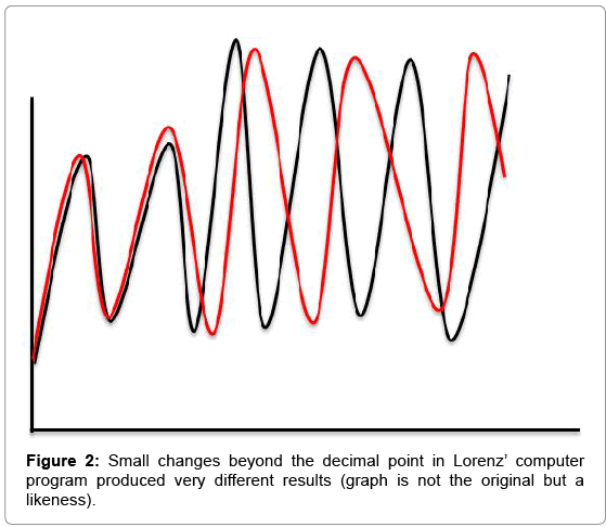
Figure 2: Small changes beyond the decimal point in Lorenz’ computer program produced very different results (graph is not the original but a likeness).
4. CASs demonstrates nonlinearity in response to perturbations.
5. CASs is composed of many components that exist at various scales. These components can be grouped into modules that communicate with each other. At lower levels of organization, some of the components are interchangeable. For example, an electron can replace another electron anywhere in the system. However, at higher levels, a module or component is unique. For example, the heart from a baboon cannot replace the heart of a human, ceteris paribus. Further, a component or module may be in several different hierarchies and respond differently to the same perturbation [81].
Alex Novikoff stated in 1947: “What are wholes on one level become parts on a higher one . . . both parts and wholes are material entities, and integration results from the interaction of parts as a consequence of their properties” [82]. Novikoff anticipated the characteristics of complex systems in noting that some systems must be studied as whole intact entities because living organisms are not “machines made of a multitude of discrete parts (physico-chemical units), removable like pistons of an engine and capable of description without regard to the system from which they are removed” [82]. The interactions of the parts are important because, according to Mayr, “a description of the isolated parts fails to convey the properties of the system as a whole. It is the organization of these parts that controls the entire system” [82].
6. A CAS is a system of systems that demonstrate redundancy of modules and robustness of the systems. For example, different genes can make the same protein through alternative splicing, thus the system will be resistant to change even though one of those genes is damaged or lost.
7. A CAS has feedback loops between components and modules, which can inhibit or amplify a response [83].
8. CASs demonstrates self-organization and are dynamic—they communicate with their environment. Communication with the environment can lead to epigenetic changes in animals and humans.
9. CASs is not simulable [84-86]. Research involving mathematical modeling is challenging this, however.
For more on the characteristics of CASs see [84-104].
I have addressed the problems with using one CAS to model another for perturbations that affect higher levels of organization and refer the reader to the following references for more on this particular aspect of CASs: [67,105-118].
The CASs I am concerned with in this article are evolved CASs. Evolution has resulted from changes in the components of the CASgenes. Genes can be mutated in numerous ways including deletions, polymorphisms such as single nucleotide polymorphisms (SNPs), and copy number variants (CNVs), Table 3 [119] for examples of gene changes influencing phenotype. All of these mutations result in changes in the initial conditions of a complex system and many such changes result in a new species. These changes in initial conditions dramatically affect outcomes to perturbations. But changes in phenotype/initial conditions can also be accomplished using other methods.
| Gene or element | Mechanism of change |
Proposed phenotype |
Phenotypic certainty |
Possible gene-associated diseases |
|---|---|---|---|---|
| AR | Deletion of regulatory DNA |
Loss of sensory vibrissae and penile spines |
Likely | Androgen insensitivity; hypospadias; muscular atrophy; prostate cancer |
| APOC1 | Pseudogene | Unknown | Not applicable |
Alzheimer’s severity; atherosclerosis; coronary heart disease |
| AQP7 | Copy number increase |
Energy use | Plausible | Nonfunctional glycerol response to exercise |
| ASPM | Positive selection | Increased brain size | Plausible | Microcephaly |
| CDK5RAP2 | Positive selection | Increased brain size | Plausible | Microcephaly |
| CCL3L1 | Novel gene variant |
Immune system function |
Likely | HIV and AIDS; Kawasaki’s disease; rheumatoid arthritis; chronic hepatitis C |
| CHRM3 | Novel exon | Change in human reproduction |
Plausible | Eagle–Barrett syndrome |
| CHRFAM7A | Copy number increase |
Higher brain function | Plausible | P50 sensory gating deficit |
| CMAH | Pseudogene | Changed sialic acid composition on all cells |
Definite | Duchenne’s muscular dystrophy; red-meat-related carcinoma risk |
| COX5A | Amino acid change |
Mitochondrial metabolism |
Plausible | Unknown |
| DRD5 | Copy number increase |
Regulation of memory; attention; movement |
Likely | DRD5 deficiency; attention-deficit hyperactivity disorder; primary cervical dystonia |
| DUF1220 and NBPF family |
Protein domain copy number increase |
Brain size | Likely | Microcephaly; macrocephaly |
| FCGR1A | Copy number increase |
Immune system function |
Plausible | IgG receptor I phagocyte deficiency |
| FSHR | Positive selection | Decreased gestation; birth timing |
Plausible | Amenorrhoea; infertility; ovarian dysgenesis type 1; ovarian hyperstimulation syndrome |
| FOXP2 | Amino acid change |
Speech and language development |
Definite | Speech and language disorder |
| GADD45G | Deletion of regulatory DNA |
Expansion of human forebrain |
Plausible | Thyroid carcinoma |
| HACNS1 | Positive selection | Changes in anterior wrist and thumb |
Likely | Unknown |
| HAR1F | Positive selection | Neocortex development |
Plausible | Unknown |
| MRC1 | Novel gene variant |
Inflammation recovery | Plausible | Leprosy manifestation |
| MCPH1 | Positive selection | Brain size | Plausible | Microcephaly |
| MYH16 | Pseudogene | Craniofacial musculature |
Plausible | Unknown |
| NCFI | Copy number increase |
Phagocyte generation of superoxides |
Likely | Chronic granulomatous disease; Williams–Beuren syndrome |
| NAIP | Copy number increase |
Inhibition of apoptosis | Likely | Spinal muscular atrophy |
| OCLN | Copy number increase |
Regulation of TGFβ; cell migration |
Likely | Hepatitis C; band-like calcification with simplified gyration and polymicrogyria |
| For references see [119] | ||||
Table 3: Partial list of genes and genetic elements showing human-lineage-specific changes (O’Bleness et al. [119]).
The regulation and expression of genes can dramatically alter phenotype and the notion that regulatory genes are responsible for major changes during evolution is now more or less universally accepted [120]. For example, gene regulation and coding area changes are responsible for stickleback evolution [121]. Polavarapu et al. studied transposable elements in humans and chimpanzees, which are thought to be important in gene regulations that were found in junk DNA [122]. One of the coauthors, McDonald, stated: “Our findings are generally consistent with the notion that the morphological and behavioral differences between humans and chimpanzees are predominately due to differences in the regulation of genes rather than to differences in the sequence of the genes themselves” [123]. Gene expression varies greatly intra- and inter-species, in humans [124-127] and in animals [128-131].
The same gene can function differently in different species. The gene Pax6 regulates brain development in humans but is apparently not required for brain development in mice or zebrafish [132,133]. Moreover, the same trait can arise through different mechanisms.
Convergent evolution is also relevant in this discussion. For example, the camera eye of humans and octopi function similarly but evolved separately and by very different mechanisms [134]. Other examples include the following:
1. Hearing in mammals developed at least twice and by different mechanisms. Monotremes and other mammals developed middle ear bones independently and by different mechanisms [135].
2. Blind mole rats resist cancer using a different mechanism from that used by naked mole rats [136].
3. Molar teeth in mammals [137].
4. Different species of bats developed different mechanisms by which they now drink nectar [138].
5. Blond hair evolved in Melanesians by a different mechanism than it did in Europeans [139].
6. Ion selectivity in neuronal signaling channels [140].
7. Clot degradation [141].
Another instrument of evolution is the old dog new trick phenomena—when an old gene is used for a new function. For example, mice, frogs, and birds use the genes Snail and Slug differently in embryonic development [145].
Genes are also influenced by background and modifier genes. Diseases such as muscular dystrophy, certain cancers, cystic fibrosis, and β-thalassemia can produce different phenotypes despite having identical gene mutations [146-148]. For example, Miklos states: “There is enormous phenotypic variation in the extent of human cancer phenotypes, even among family members inheriting the same mutation in the adenomatous polyposis coli (APC) gene believed to be causal for colon cancer” [149].
Given that all these differences among species occur in systems that are complex, and thus highly dependent on initial conditions, implies that we should expect major differences in outcomes to perturbations that occur at higher levels of organization. The importance of emergent phenomenon and the other features of a CAS are also important in how the CAS responds to perturbations and these features are also affected by changes brought about in the course of evolution.
Empirically, this has been confirmed. Efimov et al. discovered that mice and humans differ in the distribution of potassium channels in the heart [150-152]. Efimov states: “The problem is the difference in gene expression between the mouse and the human is very very large” [153]. These differences result in drugs that appear efficacious in mouse models but are ineffective in humans. The KATP channel has one of two regulatory subunits, SUR1 and SUR2 (for sulfonylurea receptor types 1 and 2) that are sensitive to ATP. In mice, the gene for SUR1 is expressed only in the atria while SUR2 is expressed only in the ventricle [151]. In humans however, drugs that bind to the SUR1 receptor do not affect the atria while drugs that bind to the SUR2 receptor affect both the atria and ventricles [150]. Efimov continues: “You can mutate in mice the gene thought to cause heart failure in humans and you don’t get the same disease, because the mouse is so different.”
Seok et al. conducted a comparative study on mice and humans measuring gene response to sepsis, burns, and trauma [154]. The background for this study was, in part, the fact that at least 150 drugs had been shown to be efficacious for sepsis in mice but all had failed in humans. Seok et al. discovered that genes expressed in these conditions varied greatly between species (Figure 3). The reaction of the scientific community is relevant to this paper. Kolata writes: “The Seok] study’s investigators tried for more than a year to publish their paper, which showed that there was no relationship between the genetic responses of mice and those of humans. They submitted it to the publications Science and Nature, hoping to reach a wide audience. It was rejected from both” [155]. Kolata then quotes RW Davis, a coauthor of the Seok article, who said: “reviewers did not point out scientific errors . . . the most common response was, ‘It has to be wrong. I don’t know why it is wrong, but it has to be wrong’” [155].
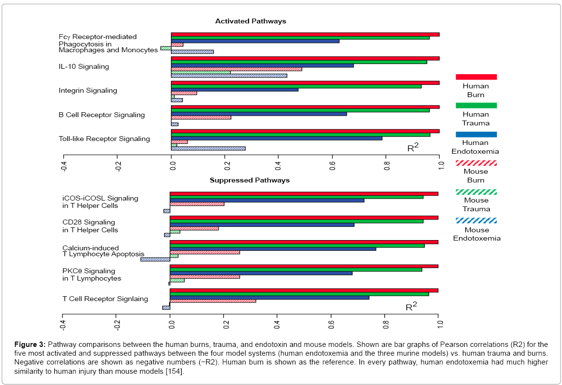
Figure 3: Pathway comparisons between the human burns, trauma, and endotoxin and mouse models. Shown are bar graphs of Pearson correlations (R2) for the five most activated and suppressed pathways between the four model systems (human endotoxemia and the three murine models) vs. human trauma and burns. Negative correlations are shown as negative numbers (−R2). Human burn is shown as the reference. In every pathway, human endotoxemia had much higher similarity to human injury than mouse models [154].
Hamlin discussed the problem that “animals used to model human diseases often have very different cardiovascular physiology from humans” [156], and noted, like Efimov, that these differences impact on the predictive value of animal models studies. Hamlin gave as an example of this phenomenon, the antihistamine terfenadine, which is cardiotoxic when combined with drugs that interfere with cytochrome P450 3A4 (CYP 3A4) [157,158]. “This effect could not be modeled in rats and mice since they do not have the hERG ion channel that caused the human arrhythmia” [156]. Multiple differences also exist between the cardiac anatomy and physiology of animals and humans [159-166].
Pfizer was forced to halt costly studies of its cholesterol-lowering drug torcetrapib due to deaths in a late-stage clinical trial [167,168]. Morgan et al. of Pfizer [169] reviewed the performance of 44 drugs from Pfizer for attrition in Phase II trials. Only 32% passed proof of concept (POC) in Phase II. They found that a majority of these drugs failed due to lack of efficacy. This is consistent with the findings of others [170-172]. They also concluded that the survival of new molecular entities (NMEs) was at its lowest during Phase II, “with small- and large-molecule survival of 38% and 53%, respectively,” which was also consistent with the data from others [173]. It is also consistent with a report from the Centre for Medicines Research that included 16 companies that represented ~60% of global R&D. The report discovered that Phase II success rates for NMEs were ~28% from 2006–2007 but were only 18% from 2008-2009 [174] of the drugs that passed POC, all had been tested in humans and “the pharmacological target was modulated as expected to elicit an effect” [169].
Suter conducted comparative research on six drugs and discovered that animals and humans shared 22 side effects but that animals incorrectly identified 48 side effects that did not, in fact, occur in humans and that animals missed 20 side effects that did occur in humans. This results in a sensitivity of 0.52 or 52% and a positive predictive value of 0.31 or 31% [175]. These values are in line with other studies and fail to qualify animal models as having predictive value.
In light of the fact that animal models are subject to Complexity Theory and the Theory of Evolution, I have developed a theory regarding the problem of using one evolved CAS as a model in order to predict responses of a second: Trans-Species Modeling Theory (TSMT) [67,105-118,176-178]. TSMT can be summarized thusly: While transspecies extrapolation is possible when perturbations concern lower levels of organization or when studying morphology and function on the gross level, one evolved complex system will not be of predictive value for another when the perturbation affects higher levels of organization” [116].
I will now analyze two advances in the treatment of cardiac disease in context of the above.
Blalock-Taussig shunt
The use of dogs in developing many surgical procedures apparently was meant to accomplish two purposes. First, to simply practice the procedure and in this the model was no doubt somewhat successful as suturing and ligating vessels is accomplished in similar ways in all mammals. Second however, was the intent to predict human outcomes both in terms of the ultimate outcome from the surgery and in an attempt to reproduce the anatomy of humans in order to ascertain whether the procedure was viable in humans. In these two endeavors, dogs performed less well. Perhaps such outcomes are what prompted René Dubos, Lasker Award winner for Basic Medical Research, to state: “Experimentation on man is usually an indispensable step in the discovery of new therapeutic procedures or drugs . . . The first surgeons who operated on the lungs, the heart, the brain were by necessity experimenting on man, since knowledge deriving from animal experimentation is never entirely applicable to the human species”.
On November 29, 1944 at Johns Hopkins University, Blalock performed the first Blalock-Taussig (BT) shunt procedure on a “blue baby” and connected the left subclavian artery to the pulmonary artery (Figure 4). The child suffered from Tetralogy of Fallot (TOF), a congenital heart disorder consisting of four features: ventricular septum defect (VSD), an overriding aorta, pulmonary outflow obstruction, and right ventricular hypertrophy due to pressure overload accompanying the pulmonary outflow obstruction [179,180]. Blalock’s lab assistant, Vivien Thomas stood behind Blalock and advised him during the surgery. The details of the operation and the development of the operation have been extensively reported elsewhere [181,182], hence I will focus on the aspects relevant to the contribution of dog models.
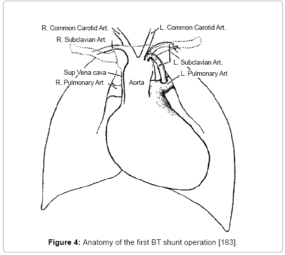
Figure 4: Anatomy of the first BT shunt operation [183].
The anatomy of the first branches of the aorta and the branches of the subclavian are similar in canines and humans. In humans the aorta branches to form the coronary arteries, the brachiocephalic trunk, left common carotid artery, and the left subclavian artery. The subclavian then branches into the vertebral artery, the internal thoracic artery, the thyrocervical trunk, the costocervical trunk and the dorsal scapular artery. In canines the branches of the subclavian include superficial cervical artery, internal thoracic artery, costocervical trunk and vertebral artery. There are intra-species variations in canines and humans, however [183-185]. Moreover, the relative lengths of the vessels and their relationships to each other may vary from species to species and individual to individual. As we will see, Blalock used the innominate artery for patients two and three instead of the subclavian because the anatomy favored that technique.
The story of the BT shunt centers on Helen Taussig, Alfred Blalock, and Vivien Thomas. Taussig was a cardiologist who had conducted many autopsies on children who died because they were “blue babies.” She explored the anatomy and decided that such babies did better if they had a persistent or patent ductus arteriosus (PDA). Taussig originally approached Gross regarding creating shunt to allow more blood to flow to the lungs because she knew that Gross had ligated a PDA [186]. This was the event that led her to ponder whether one could also create such a duct [187]. Taussig relied on clinical observation of patients along with autopsies to formulate a hypothesis regarding treatment [183].
Alfred Blalock had met Vivien Thomas, a black man who aspired to attend medical school at Vanderbilt University in the 1930s. Thomas’ hopes were dashed because of financial woes and this led him to apply for a job as a lab technician. Thomas’ story is told in the movie something the Lord Made and in his book Partners of the Heart: Vivien Thomas and His Work with Alfred Blalock: An Autobiography. In 1938, Blalock and Thomas were at Vanderbilt attempting to induce pulmonary hypertension in dogs by performing an end-to-end anastomosis of the subclavian artery and a branch of the pulmonary artery [188]. They apparently failed in their attempt but this would eventually be the very procedure Blalock would perform on blue babies. Thomas accompanied Blalock to Baltimore when Blalock accepted the position as surgical chief at Johns Hopkins University.
After Taussig failed to convince Gross to attempt to create a surgical duct, she contacted Blalock. Blalock and Thomas subsequently attempted to create a dog model of the pulmonic stenosis aspect of TOF [183]. They did not attempt to reproduce all of the anomalies of TOF, just the outflow obstruction and the limited blood flow to the lungs. They did reproduce one of the symptoms of TOF-cyanosiseven though it was by a different mechanism from TOF. In fact, the way they accomplished the cyanosis had little in common with the actual anomaly in blue babies. Blalock and Thomas removed a portion the dog’s lung and created a pulmonary arteriovenous fistula. This resulted in cyanotic dogs. They then connected a systemic artery to the pulmonary artery and thus restored normal blood flow to the lungs. This resulted in the dogs being well oxygenated again. Note that the dog model really did not mimic human TOF or pulmonary atresia, or any heart anomaly that resulted in blue babies. This was not a deterrent to testing Taussig’s hypothesis however, because her hypothesis revolved around plumbing and flow, which in physics is described as fluid dynamics [181,182] or even Ohm’s law, modified for flow: flow ☒(pressure / resistance). Simple physics dictates that providing more flow through pipes designed with the capacity to carry greater volume will result in more volume being delivered, ceteris paribus, and this is the case for the BT shunt [189-191]. This fact alone calls into question the necessity of the dog model in the development of the BT shunt. Moreover, as I noted in the introduction, flow increase via the EC-IC bypass procedure was also intuitive, worked well in dogs, but failed in humans. While the laws of physics do not vary from species to species, physiology does and hence positive outcomes in animal models are not predictive for humans.
Three other systemic-to-pulmonary shunts were also developed shortly after the BT shunt: the Potts anastomosis, the Glenn shunt, and the Waterston anastomosis [192-194]. All could have been intuitively derived based on fluid dynamics. Interestingly both the Waterston and Potts have been abandoned due to a flow rate that is too high [195]. Such was not anticipated from the dogs studies performed prior to implementing the procedures in humans. The Glenn shunt was also abandoned due to complications not observed in the original dog studies [196,197]. Similarly, the major complications from the BT shunt, such as impaired left upper limb development, were not anticipated from dog studies [183,195,198]. This reinforces what I have previously stated regarding perturbations that affect conserved processes or processes primarily governed by the laws of physics: even when an effect is conserved the side effects are unique [114].
Gross et al. had anastomosed the subclavian artery to the pulmonary artery in animals prior to 1941, when Blalock accepted the chairmanship of surgery at Johns Hopkins [199]. Gross et al. concluded that this had increased blood flow to the lungs and thus exposed more unsaturated blood to the alveoli. As previously mentioned, Blalock had published a paper describing subclavian anastomosis in 1939 [188]. It is odd that few of the authors recounting the history of the BT shunt have mentioned this, as it is the exact operation Blalock performed. Regardless of past experiences, using the subclavian artery to increase blood flow to the lungs of blue babies was Taussig’s idea [200]. All of this again casts doubt on the value of the dog model as used by Blalock and Thomas after Taussig consulted with them regarding what would become the BT shunt procedure. Granted, it simply moves the dates back, in term of the importance of dog models, to Blalock and Gross’ previous surgeries of the 1930s that had already established that subclavian artery to pulmonary artery anastomoses could be performed in the dog. This backdating of the surgeries, however, is relevant for this discussion as the claim made by proponents of the position that the dogs operated on by Thomas, after Taussig had suggested the subclavian shunt, were necessary for the operation to be performed in humans. For example, Murphy and Cameron state: “Using this animal model Blalock’s team demonstrated that anastomosis of a systemic artery to the pulmonary artery was feasible and improved the arterial oxygen saturation” [201]. This lack of attention to detail does not bode well for the claims of animal modelers in general or the claimed importance of the dog studies at Johns Hopkins.
Furthermore, Blalock never performed the shunt surgery on any of the 200 dogs Thomas operated on at Johns Hopkins; he merely assisted Thomas on one dog [181,182,187]. History does not appear to record whether Blalock operated on any of the dogs at Vanderbilt that were used in the pulmonary hypertension experiments. Given Blalock’s avoidance of the dog lab at Johns Hopkins for a procedure that he was supposed to perform, one can reasonably assume he never operated on any of the dogs at Vanderbilt since he was not anticipating performing the operation at Vanderbilt. Cooper confirms that Blalock had not performed the procedure in the Johns Hopkins dog lab prior to trying it a patient [26]. Blalock stated that he wanted to practice the surgery prior to performing it, but Taussig’s first candidate for the procedure was deteriorating and the surgery was an emergency. Hence Blalock did not have time to practice the procedure.
Based on the above, I do not think the claim by the proponents of the importance of the operations Thomas performed on dogs at Johns Hopkins can be taken seriously. For knowledge transfer to occur between Thomas and Blalock, one would have to appeal to metaphysics. Merely watching a technician perform a surgery on a dog and having that technician standing behind the surgeon during the procedure on a human would not be of any benefit for the patient. If it were, surgical residencies would dramatically modify their teaching methods. Ideally, a surgeon will observe the procedure being performed on patients many times before attempting pieces of it depending on his level of training. Even experienced surgeons watch a new procedure in person or on video prior to performing it. No one watches a new procedure performed on a dog and thinks himself qualified. However, if Blalock were, in reality, seeking data on the mechanism of the cyanosis and attempting to reproduce that in the dogs, already knowing that he could suture an end-to-end anastomosis, then he would feel under no pressure to practice on dogs.
Likewise, even in the case of new surgeries performed on an emergency basis, the surgeon is more likely to benefit from the experience he has and his knowledge of the anatomy and surgical technique in general. Many new surgeries have been successfully performed, emergently, for example in battlefield scenarios or even in homes, such as Ephraim McDowell’s oophorectomy in 1809 [202]. Surgical expertise in general has figured largely in the outcomes. The claim that knowledge transfer occurred between Blalock and Thomas based on all the surgeries that Thomas performed at Johns Hopkins does not stand up to scrutiny. There may be another reason however, why Blalock had Thomas in the operating room, however. Cooper acknowledges, as do others [62], that Blalock was not a technically talented surgeon. He may have had Thomas there for moral support. Given that many procedures were not attempted due to irrational biases, having Thomas there may have been a security blanket for Blalock. Regardless, given Blalock’s experience as a surgeon and the relatively straightforward nature of the anastomosis, advice from a technician hardly seems warranted.
The above also forces one to reconsider the importance of the dog procedures at Vanderbilt [188]. Proponents of the importance of the dog model for the BT procedure should cite those operations as being necessary if any operations on dogs were necessary for the attempt in humans. But what did even those procedures really accomplish? Blalock and Thomas were attempting to create a model for pulmonary hypertension; the creation of a shunt was a secondary concern. In his 1939 paper describing those operations, Blalock emphasizes the idea to create pulmonary hypertension not the anastomosis, which is described in one sentence on the first page of the article. If there was a prohibition or tale of caution warning against ligating and anastomosing the subclavian artery, no mention is made in Blalock’s papers. Moreover, such an admonition would have been irrational in retrospect. Many surgeons have gained confidence from performing new procedures on dogs only to see a very high mortality rate in their first patients. Granted, some of the new procedures worked well, but given the ones that did not, the predictive value of such experiences must be called into question.
Finally, Thomas’ experience with operating on dogs had left very important questions unanswered. Unfortunately, the first patient to have a Blalock-Taussig procedure died less than one year after the operation probably secondary to a failure of the surgical procedure to last a sufficient period of time. The importance of practicing the shunt on dogs is also called into question as the innominate artery to pulmonary artery anastomosis was not practiced, and yet in the second and third patients to receive the operation, Blalock connected the innominate artery to the pulmonary artery instead of the subclavian because of the patients’ anatomy.
Another experience at Johns Hopkins that predated Blalock and Thomas’ experiments with dogs because of the consultation with Taussig was an attempt to create and correct a coarctation of the aorta. Inspired by Edwards Parks, Blalock and Thomas created a coarctation and then attempted to repair it by performing an end-toend anastomosis of one of the carotid arteries or the subclavian artery. The dogs responded poorly and Blalock abandoned the idea [187,203]. Here we again see another instance of an end-to-end anastomosis that involved the subclavian artery being performed in dogs.
Though the first BT shunt procedure was initially a success, the baby girl that Blalock operated on became cyanotic a few months later. A second procedure was performed but she did not survive. It would later be discovered that children over three years of age were more likely to survive the BT shunt procedure [204]. The use of synthetic shunts eventually replaced the subclavian or innominate artery as the exact length can be determined intraoperatively, limb perfusion is not compromised, and less dissection is required to place the shunt, among other factors [205].
This analysis is not meant to disparage any of the three main people that participated in the development of the BT shunt. Thomas was obviously brilliant and made many contributions to medicine including the invention of new surgical tools for the BT shunt and vascular surgery in general. Vascular surgery was not common at this time and the specialized clamps and needles used today were not available [181,182]. The reputations of Taussig and Blalock speak for themselves. This section addressed the claim, made by current scientists who uncritically advocate for the use of animal models, that the operations on dogs that were performed at Johns Hopkins were necessary for the development of the BT shunt operation. I will address the implications of these specific claims later in this article.
Intra-arterial stents
Heart disease (HD) is the leading cause of death in developed nations and is increasing in incidence as developing nations adopt Western lifestyles. In the US, HD is the number one cause of death in both men and women, causing over 600,000 deaths in 2008. One out of every four deaths in 2008 was attributed to HD. Coronary artery disease (CAD) is the most common type of HD, causing over 400,000 deaths in 2008 [206]. Over one million people experience a heart attack each year in the US [207]. CAD is estimated to have cost the US economy over $1 billion in 2010 [208]. The introduction of stents for the treatments of CAD and acute myocardial infarction has revolutionized health care. The placement of intracoronary stents (Figure 5) is the most commonly performed therapeutic procedure in medicine [209] with over 500,000 patients undergoing the procedure in the US annually [207]. I will begin the analysis of the role of animal models in stent development with a brief history of invasive cardiac procedures.
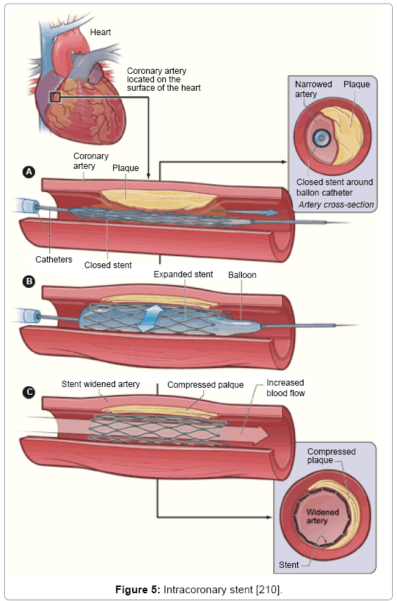
Figure 5: Intracoronary stent [210].
According to Cooper [26], Galen, of the second century CE, learned much about the heart from attending to gladiators who expired from chest wounds. Cooper also comments on the fact that much of what Galen learned from animals turned out to be wrong in terms of human anatomy. Despite these inaccuracies, the Holy Roman Church accepted Galen’s discoveries and anyone disagreeing with them was subject to execution. One such unfortunate was Miguel Serveto, who pointed out that Galen was wrong on some aspects of circulation only to be burned at the stake for his efforts [26]. William Harvey of the 17th century demonstrated that blood was circulated through the body by the heart. Horses and frogs were used in this endeavor. I note that animal models of the heart and circulation in general were used to gain insights into the function and gross anatomy of these systems. This is consistent with categories 5 and 6 in Table 1.
In 1711, Stephen Hales successfully placed a crude catheter into both the right and left ventricles of a living horse [210,211]. Claude Bernard also catheterized the hearts of animals in the 19th century [212]. However the first successful human heart catheterization was self-performed by Werner Forssmann, in 1929 [213]. After inserting the catheter into his antecubital vein, Forssmann walked up a flight of stairs and confirmed placement with an x-ray of his chest. Cournand and Richards used Forssmann’s technique to measure the hemodynamics [214], and they, along with Forssmann were awarded the Nobel Prize in Physiology or Medicine in 1956. Animals such as dogs could have been, and no doubt were, used to practice heart catheterization and to obtain the data necessary to calculate hemodynamic values. However, the literature seems to indicate that humans were the subjects of choice for most of these endeavors. Catheterization and visualization of the left human heart would not occur until 1953 [215]. Regardless, using animals to discover basic physiological principles (number 5 in Table 1) and to learn the basics regarding how to perform simple procedures (number 6 in Table 1) are viable uses of animals and are separate from categories 1 and 2 in Table 1.
In 1927, Portuguese physician and future Nobel laureate Egas Moniz of the University of Lisbon, was the first to develop angiography, specifically cerebral angiography. Moniz developed the procedure on dogs, monkeys, and human cadavers using strontium bromide, but when injected in living humans the first patient in whom Moniz achieved vascular visualization died. Moniz continued his efforts and eventually succeeded in visualizing the cerebral vascular system using sodium iodide [216-218]. Sodium iodide had also previously been studied in cadavers. Various chemicals were used in an attempt to find a contrast agent that was safe and effective. For example, intraarterial administration of Lipiodol was associated with death in dogs but was apparently safe and effective in humans [219]. Dandy had already developed ventriculography but it had a 10% mortality rate along with limited visualization [220]. Angiography was also aided by the development of the radiocarrousel by the radiologist Caldas, which allowed numerous radiographs to be made over several seconds [220]. The use of animals to study safety and efficacy of the dyes is an example of using animals as predictive models for humans and apparently was not viable for Moniz. Safe and effective dyes were eventually found essentially by trial and error.
The radiologist Charles Dotter was responsible for several advances in percutaneous cardiac intervention including the doublelumen balloon catheter and the guidewire [221,222]. Dotter’s most important contribution however was the angioplasty. He first placed a coil-spring stent in the femoral artery of a dog and later in humans. Dotter’s contributions reflected his interest in engineering, which was manifest even as a child. Although Dotter experimented on dogs, his contributions were of an engineering nature and the role of dogs appears to have been nonessential. Also of interest is the fact that Dotter’s first recanalization was an accident. In his attempt to perform an aortogram of the abdominal aorta, Dotter passed the catheter through the occluded right iliac artery [223]. Dotter also pioneered a technique to visualize the coronary arteries by occluding the aorta momentarily and injecting dye into the aortic root, a technique he likewise practiced on dogs [224]. Interestingly, Dotter did not claim to be the first to visualize the coronary arteries via angiogram. Various attempts had been periodically made and the first success appears to have been in 1933 [225,226]. Other successes followed, perhaps greater than 20, both in humans and animals using various techniques and with varying degrees of clarity of visualization [224]. Whenever a procedure involves basic physiological principles or can be described in terms of a simple system, animal models will likely provide insight. (For more on this concept see [114].) As history does not record the specifics of many of these advances, it is difficult to determine when animal models were necessary, redundant, or misleading.
In 1958, pediatric cardiologist Sones accidently demonstrated opacification of the right coronary artery while attempting ventriculography [55,227]. This technique of direct injection of a coronary artery supplanted Dotter’s procedure. At this time, injection of contrast into the coronary artery was thought to produce ventricular fibrillation and certain death [26]. Sones’ accident changed the course of interventional cardiology. The importance of the dog model in coronary angiography is severely undermined in light of the fact that the current practice of coronary angiography is based on an accident in a procedure performed on a human.
In 1974, Gruentzig placed a balloon on the end of a catheter and thus enabled physicians to dilate an artery occluded by plaque [228,229]. He practiced the technique in dogs and performed the first percutaneous transluminal coronary angioplasty (PTCA) in 1977 [228,230]. The use of wires to guide the catheter as well as the attachment of a balloon resulted from advances in engineering.
Five years later, in 1979, Rentrop placed a catheter into the left anterior descending coronary artery and injected streptokinase in an attempt to prevent a myocardial infarction [231]. Streptokinase had been injected systemically in previous attempts but patients suffered from severe hemorrhage as a result. The direct injection using less streptokinase proved efficacious in addition to limiting the side effects [231]. Streptokinase is a product of the metabolism of hemolytic streptococcus and was first proposed as a clot buster in the early 1950s [232-235]. Experiments on animals revealed varying effectiveness due to species variability [236]. Alkjaersig et al. stated: “Biochemically, considerable species differences exist not only between the plasminogen system of man and animals, but more particularly between the systems of various animals; this variability is most extreme with regard to the differential effectiveness of streptokinase” [236]. Nevertheless, intravenous injection of streptokinase did result in dissolution of thrombus at least some of the time in animals.
Experiments in humans soon followed with streptokinase also proving able to dissolve clots. However, administration was accompanied by fever and hypotension, as the samples of streptokinase were not pure [235,237-239]. With purification, these side effects disappeared [237,240]. Much of the biochemistry regarding clots and clot dissolution was worked out using in vitro methods [236]. By the end of the 1950s the safety and efficacy of streptokinase administration for acute myocardial infarction in humans had been established [240] with more clinical trials ongoing. Eventually intracoronary injection became the preferred method of administration [231,241- 244]. The administration of anti-coagulants was also important in the development of stents but, like streptokinase, also caused severe side effects in addition to aiding in the patency of the vessel.
Ulbricht and Southgate stated the following regarding the development of drugs, such as rt-PA, to treat thrombosis: “Some of the early optimism for rt-PA as a thrombolytic agent was based on experiments conducted in animal models of thrombosis. The clot specificity of rt-PA in animal models was far more pronounced than that observed in subsequent clinical experience in man. This is in part due to the non-occlusive nature of the animal models of thrombosis used and the poor activation of animal plasminogen by the nonhomologous human rt-PA” [245].
The claim that animal models were necessary for stent development appears to rest on three separate claims. First that animal models can predict safety, or lack of toxicity, of the stents and the drugs used in the stents. Second, those animal models can predict stent efficacy. Third, claims that animal models were necessary for medical advances have also relied on discoveries in the distant past, such as discoveries regarding the fundamentals of physiology. Although the claims regarding stent development have not always included historical references, I nevertheless addressed some of them above. As I stated previously, due to the fact that many of the experiments and advances were not analyzed at the time nor were the exact details recorded, it is difficult to ascertain where animal models were necessary. Some of the advances were merely applications of some of the basic principles of physics, while, in other cases, animal models demonstrated effects but not side effects. In reading the cardiac literature, one cannot help but be affected by how high the human mortality rates were after a procedure had been perfected in the dog lab.
After the development of coronary angiography and thrombolytic therapy, physicians considered placing a stent in the coronary artery in order to prevent occlusion or re-occlusion. Dotter and Judkins suggested the notion of using stents in arteries in 1964 [221]. After completing the procedure in dogs, Puel et al. were the first to place stents in humans in 1986. In 1989, they used a stent after balloon angioplasty in order to prevent re-occlusion. Although restenosis and occlusion were not seen in the animals studied by Puel et al, the complications were observed in humans [246-248]. These first stents were bare metal stents (BMS) and unfortunately were associated with a high incidence of subacute thrombosis in humans.
Palmaz et al. first employed stents in peripheral arteries in 1985 [249,250]. This stent was subsequently modified to the Palmaz–Schatz stent, a heparin-coated stent [251-253]. The heparin-coated stent was superior to the BMS [254] but would ultimately be replaced by the drug eluting stent (DES). The first DES was Cypher, introduced in 1995, which released sirolimus. The Taxus stent was next and released paclitaxel. Both drugs interfere with mitosis. The DES has been consistently shown superior to the BMS [255-257].
The DES consists of a stent, a drug to inhibit restenosis, and a method for delivering or releasing the drug. The physical structure of the stent has evolved based on advances in engineering. Originally, stainless steel was favored but currently metal alloys are being used. The alloys allow for thinner struts, which allow faster endothelialization and less injury to the vessel. Stents can vary in length, the thickness of the struts, and the alloy. These variables affect the radial strength, flexability, radiopacity and recoil of the stent. Current alloys include cobalt–chrome and platinum–chrome. A stent should have the following qualities:
• Mechanical resistance to abrasion during implantation
• Suitable for sterilization
• Allow time- and dose-controlled drug release
• Suppress thrombogenesis and inflammation of the vessel wall and tissue [258].
Mechanisms for drug release have also evolved with polymer coatings being used most frequently [259-261]. Various combinations of polymers allow for different rates of diffusion of the drug [262]. These are examples of advances in engineering and I found no claims that they were due to animal models.
The sirolimus stent was a success in humans as was the paclitaxeleluting stent [258], however this was not what animal studies suggested (Unpublished studies from AJ Carter in 2002 and AW Heldman in 2002 as cited in [263] and [264]). Studies in pigs revealed no benefit from these stents at three and six months. This led some to question the predictive value of animal models for stents [265]. This phenomenon repeated itself with brachytherapy. No long-term benefit was seen in animal models but humans did benefit [263,265-269]. Serruys et al. state: “Finally, because the results of experiments in animal models cannot be directly translated to humans, specific clinical trials of safety and efficacy are required for each device [DES] [270].
Intracoronary stents prevent the artery from occluding and hence prevented the patient from experiencing a myocardial infarction (MI). This is simple physics. But the stents also led to the problem of subacute thrombosis and in-stent neointimal hyperplasia from scar tissue growth-intimal proliferation [271]. Despite the failure of animal models referred to above, Perkins states: “Because of the complex, multidisciplinary, and dynamic nature of this technology, thorough evaluation of DES systems in preclinical models is crucial for predicting clinical safety and efficacy as well as for providing details into the pathophysiology of the vascular response to injury and restenosis” [272]. Clearly Perkins is claiming that animal models have predictive value. This claim is not unique to Perkins.
The safety claim fails, in part, due to the fact that the drugs being used have been administered to humans for years and the toxicity profiles are well known. The second component of the DES is the metal component and while metal allergies exist, allergies and adverse reactions to metal alloys appear to be rare and idiosyncratic [273,274]. In summary, there is nothing new in terms of traditional toxicity to be learned from the implantation of DESs in animal models, thus claiming that animal models are predictive for human toxicity is an example of the fallacy known as argument by half-truth. It is true that DESs have not resulted in toxicities such as hepatotoxicity, but the reason has nothing to do with animal models and is, in actuality, due to the fact that the drugs have been used in humans for years. It is also an example of post hoc ergo propter hoc. The novel toxicity would be intimal hyperplasia, which I will discuss below.
Numerous species have been models for restenosis in humans. These include dogs, nonhuman primates, rabbits, rodents, sheep, and swine [272]. None of the models demonstrates the underlying atherosclerosis of humans. Perkins continues stating that murine models are easy to handle, inexpensive to maintain and house, can be studied reliably in high volumes, and reveal a number of molecular markers. Unfortunately, none of these characteristics are relevant to the issue of predictive value. Perkins also states that as these models do not exhibit atherosclerosis, it must be induced in some fashion and thus: “These models have limited application: There is little thrombus formation, and the induced neointimal hyperplasia tends to be smooth muscle cell rich with little resemblance to human pathology specimens” [272]. Of the pig model for restenosis, Perkins states: “The neointimal response is of a similar histology to that in restenotic human coronary arteries; however, as with other preclinical models, the degree of restenosis often is not sufficient to be of clinical significance even with heightened injury through overstretch models” [272]. Pigs also develop and eosinophilic inflammation in response to basically all stent implantation [272]. Dogs, sheep and NHPs also demonstrate significant difference from humans in terms of stent response [272]. All of this calls into question Perkins’ claim of predictive value for animal models of intra-arterial stents.
In fact, animal models have consistently misled regarding restenosis and occlusion. For example, studies in dogs suggested that coating the stent in gold would decrease restenosis [275,276] but when studied in humans the gold increased the rate of restenosis [277]. Conversely, when sirolimus-eluting stents were studied in pigs, the factors thought to contribute to restenosis were favorably influenced but only for a limited period of time. Neointimal area was less than controls at 30 days post-implant but greater than controls at 90 days post-implant [270]. The authors of the study stated: “The clinical efficacy of SRLeluting stents would not be expected based on the degree and duration of suppression of neointimal formation documented in normal porcine coronary arteries. The vastly different pharmacodynamics of SRL-eluting stents observed to date in human clinical trials versus preclinical models may be attributed to differences in species response to SRL, anatomic substrate and physiological stimulus for neointimal formation” [270].
The rat carotid artery model revealed that angiotensin-converting enzyme (ACE) inhibitors prevented or retarded neointimal thickening [278,279]. Human studies failed to replicate these results, however [280,281]. The rat model also failed to mimic other human responses [282-284]. Sprague states that the rat carotid injury model “exhibits intimal smooth muscle cell hyperplasia similar to the human arterial response to balloon angioplasty-induced injury but does not exhibit mural thrombosis or inflammation at the injury sites, as commonly observed in humans” [285]. These are not insignificant differences.
Administering systemic medication to prevent restenosis failed in humans despite favorable outcomes in animals [286-292]. De Feyter, Vos, and Rensing state: “Recent randomized trials using fraxiparine (an interventional agent), pravastatin, nitric oxide donors, fluvastatin, octreotide (a somatostatin analog), supplementation with omega-3 fatty acids, carvedilol, or amlodipine did not demonstrate a reduction in either restenosis nor target vessel revascularization when compared with placebo. This was rather disappointing because in animal experiments these pharmacologic agents were invariably positive, whereas in the human clinical setting they were not effective” [293]. In 65 trials involving over 25,000 patients no drugs were shown to be both safe and effective in preventing restenosis despite positive results in animal models [265,293-296].
Compare the above results with the following statement from Schwartz, Chronos, and Virmani: “The examination of neointimal hyperplasia in injured animal artery models has helped in our understanding of angioplasty and stenting mechanisms, and as drugeluting stent (DES) technologies have arrived, they too have been advanced through the study of animal models. These models are useful for predicting adverse clinical outcomes in patients with DESs because suboptimal animal model studies typically lead to problematic human trials. Similarly, stent thrombosis in animal models suggests stent thrombogenicity in human patients. Equivocal animal model results at six or nine months occasionally have been mirrored by excellent clinical outcomes in patients. The causes of such disparities are unclear but may result from differing methods, including less injury severity than originally described in the models. Ongoing research into animal models will reconcile apparent differences with clinical trials and advance our understanding of how to apply animal models to clinical stenting in the era of DESs” [297]; emphasis added.)
Schwartz et al. clearly state that the porcine model is predictive for stent thrombosis [297] but the basis for this appears to be isolated instances of correlation rather than a series of comparisons which would be required for calculating PPV and NPV. Most effects from DES have been demonstrated in an animal models but no model has consistently shown itself to have predictive value for humans. Moreover, retrospectively finding an animal model that reacts to sirolimus eluting and paclitaxel eluting stents as humans react is not consistent with the scientific definition of predictive. In order to calculate the predictive value of the model, all the successes and failures of the model must be known and those values plugged into the formulas in Table 2. The reality is even worse however as animal models revealed that the sirolimus eluting and paclitaxel eluting stents were ineffective at six months while humans responded much more positively [263,285]. One must question whether such data from animals caused drugs for DESs that would have been effective for humans to be discarded [298,299]. I encountered no calculation of PPV and NPV of animal models for stent assessment despite many proclamations of the predictive value of such models [263,265,300,301]. For example, Virmani et al.: “It can be argued that insufficient preclinical testing may have led to the recent failures of actinomycin-D (European Society of Cardiology Congress 2002, unpublished data) and the paclitaxel derivate (QP2 or 7-hexanoyltaxol) eluting polymer stents in de novo and restenotic lesions. It should be recognized that the vast knowledge of vascular healing and repair derived from animal studies is echoed in today’s clinical achievements in the field of stent restenosis” [263]. Given the data I have presented, it is highly unlikely that animal models have obtained a PPV and NPV high enough to be considered of predictive value.
Neither small nor large animal models have been predictive for interventions to prevent restenosis [302]. Likewise, both have been unsuccessful for simulating the lesions of atherosclerosis and restenosis [302]. Johnson et al. state: “Some of the reasons for the frequent use of small animal models in restenosis research include, a) low cost, b) ready availability, c) reduced ethical concern compared to large animals-especially primates-and d) small size that limits the quantities of new agents required for in vivo screening. These characteristics have permitted rapid evaluation of new agents in sufficiently large populations to perform meaningful statistical analyses. In addition to these practical indications for their use, small animal models have the added advantage of well-defined genetic characterization, and, in the case of mice, the availability of transgenic and gene knocks-out animals.” [302].
The above reasoning is interesting, but again is not relevant to predictive value. Further, Johnson continues: “Despite the favorable characteristics of small animal models, the predictive value of the data obtained from the study of small animal models has been very limited. Greater than 40 large-scale clinical trials, that included thousands of patients, failed to establish significant effectiveness of multiple pharmacological agents in the prevention of restenosis following human angioplasty, even though most of the agents evaluated had been found to reduce luminal narrowing following arterial injury in small animal models. Only recently have a limited number of interventions been shown to reduce the rate of restenosis following angioplasty in humans. Examples of several drugs that have been shown to inhibit intimal hyperplasia and/or luminal narrowing of small animal arteries following angioplasty or other forms of arterial injury, but that have failed to influence coronary restenosis in humans, are shown in the Table 4 along with the three agents – probucol, triazolopyrimidine (trapidil) and irradiation – that have reduced restenosis rates in clinical trials” [302]. A perusal of Table 4 will reveal that it is difficult to calculate PPV and NPV for, among other reason, the fact that the same species have opposite results in many cases. This brings us back to personalized medicine and the fact that even human respond differently to drugs and disease. Regardless, there is insufficient data to conclude any species has predictive value but sufficient data to conclude that no species has been proven to have predictive value for human response. Given TSMT, there is no reason to assume animal models will ever have predictive value for perturbations that occur at higher levels of organization, such as response to drugs and disease.
| Intervention | Results | ||
|---|---|---|---|
| Small Animals | Large animals | Humans | |
| Heparin | Rat + Rabbit + Rabbit - |
- | |
| LMW Heparin | Rabbit + | Pig + Baboon - |
- |
| Dipyridamole +aspirin | Rabbit + Rabbit + Rabbit - |
- | |
| Ticlopidine +aspirin | Rat – Rabbit - |
- - | |
| Prostacyclin | Rabbit + | Pig + Pig - |
- |
| Glycoprotein IIb-IIIa Inhibitors | Hamster + Rabbit - |
abciximab ? eptifibatide ? tirofiban - |
|
| Aspirin | Rat – Rabbit - |
Pig + | ? - - |
| Thromboxane Receptor Antagonists +Ketanserin | Rabbit - | Dog + | - |
| Calcium Channel Blockers | Rat + Rat - |
- | |
| Corticosteroids | Rabbit + | - | |
| ACE and Angiotensin II Inhibitors | Rat + Rat – Guinea Pig + Rabbit + |
Pig – Baboon - |
+ - |
| Statins Lovastatin Fluvastatin Pravastatin |
Rat + Rabbit + |
+ - - - |
|
| Hirudin | Rat + Rabbit + |
Pig + Pig - |
- |
| Somatostatin Analogs Angiopeptin Octreotide |
Rat + Rabbit + |
Pig + | -- |
| Cell Cycle Inhibitors | Rat + Rabbit + |
Pig + Pig - |
- |
| Cilostazol | Rat + | Dog + | + |
| Irradiation | Rabbit + | Pig + | + |
| Trapidil | Rat + Rabbit + |
+ | |
| Antioxidants Probucol Others |
Rabbit + Rabbit + |
Pig + | + |
| + Inhibited intimal hyperplasia and/or luminal narrowing in animals or restenosis in humans | |||
| - Inhibited intimal hyperplasia and/or luminal narrowing in animals or restenosis in humans | |||
| ? Data insufficient to determine | |||
Table 4: Effect of interventions to inhibit arterial narrowing in animal models and restenosis in humans [302].
Yet, scientists continue to hype animal models for increasing the probability for safe and effective interventions. For example, Schwartz et al., who stated above that animal models are predictive, also stated the following: “it is unclear that any single animal species is more indicative of the potential human clinical response and for the indications desired. As such, animal models provide mechanistic insight into fundamental biological processes and appear at a minimum to indicate relative safety. Furthermore, preclinical models allow testing critical hypotheses regarding putative mechanism of action of an intervention. There is no perfect animal model of human vascular disease. Research into correlative data between animal models and human clinical application is underway in hopes of predicting therapeutic features of safety, efficacy, and practicality in reliable animal models. Proof of concept can be examined in animals including evidence for toxicity based on histopathologic effects and advanced cell/tissue analytic techniques. True efficacy and safety can currently only be proven in humans, so it is critical to construct human trials that resemble the animal preclinical trials and to make it clear what data and important conclusions can be justifiably extracted from animal models” [303].
This statement contains many fallacies and reveals a lack of understanding of the fundamentals of the philosophy of science. If no “single animal species is more indicative of the potential human clinical response and for the indications desired” than any other species, then animal models fail to have predictive value. While it is true that animal models “provide mechanistic insight into fundamental biological processes”-for example the Krebs cycle is more or less the same across species lines [304,305]-the response to stents is not a fundamental biological process. Safety relates to the nontoxicity of the drugs and alloys used in the stent and, as drugs and chemicals have been extensively studied in animal models and the animal models shown to fail as predictive modalities, the safety of stents cannot be assured after animal studies. Moreover, as animal models fail to be of predictive value for outcomes, it is nonsensical to believe that they “allow testing critical hypotheses regarding putative mechanism of action of an intervention.” I will allow the rest of the statement from Schwartz et al. to speak for itself. Sprague’s statement is closer to reality: “The complete validity of any of the specific animal models for evaluating the various drug therapies which target atherosclerosis or angioplasty-associated restenosis can only be determined after extensive clinical experience. This is evident as clinical studies continue to examine the extent of late thrombosis associated with the use of drug-eluting stents” [285]. Hau anticipated this year ago: “It is not possible to give reliable general rules for the validity of extrapolation from one species to another. This has to be assessed individually for each experiment and can often only be verified after first trials in the target species” [306].
Scientists who use animals as CAMs assume that a general similarity in a subsystem, such as response to neointimal injury, implies causally related mechanisms and causally related responses to other perturbations such as stent placement. Virmani et al. state: “In animals or humans, the local response to a bare stainless steel stent in normal or diseased atherosclerotic coronary arteries follows a distinct pattern of arterial injury and repair accompanied by some degree of neointimal formation and endothelialisation. These healing events, however, can be notably altered with the addition of polymers, antirestenotic drugs, or both” [263]. In this assessment, Virmani et al. are noting similarities between animals and humans and offering this as a basis for employing animals as CAMs for humans. However, Virmani et al. continue by stating: “Nevertheless, it is poorly appreciated that neointimal responses are exaggerated and that the time course of healing is more prolonged in humans than in animals” [263]. This statement references empirical evidence revealing no causal relationship between outcomes of stent placement in animal models and in humans. Moreover, as I explained above, scientists should not expect causal relationships between subsystems of intact evolved systems that are differently complex.
Ramifications of attributing BT and stents to animal models
Clearly, there are things medical science can learn from studying animals (I have discussed this in reference [114]). Properties subject to the laws of physics, for example, will probably result in similar outcomes across species lines. Fluid dynamics will be the same regardless of the species under study. Likewise, suturing an endto- end arterial anastomosis will demand more or less the identical skills regardless of species. Making a small arterial opening larger can correspondingly be accomplished in many of the same ways across species lines. Physiologic response to the above varies however. This knowledge would be classified as trivial for any biomedical scientist. The reasons animal models cannot be CAMs for responses that occur at higher levels of organization, and hence the reasons why animal models cannot be of predictive value, lie in the dissimilarities among species via the changes produced by evolution.
The length of artery necessary to supply a new area of the body must be determined in vivo in the individual in question. Cadavers can give one an idea of whether the subclavian artery can be used as a shunt but individuals vary as exemplified by Blalock’s choice of the innominate artery in patients 2 and 3. Dogs have their longitudinal axis parallel to the ground while humans are perpendicular. We should expect some variation because of this although how much is uncertain. Moreover, whether the variation is clinically significant can only be determined by comparing the trait in question, for example length of the subclavian artery, among species. Practicing arterial anastomoses in dogs offers little to the surgeon who already knows how to create an arterial anastomosis.
The BT shunt is performed for dogs and other animals with TOF but it must be modified from the original as the subclavian does not have sufficient length to connect to the pulmonary artery without kinking at the origin [307]. TOF can also be corrected in dogs in more or less the same manner it is corrected in humans [307,308]. This is due to the fact that the nature of the pathology, and hence the correction, is based on fluid dynamics.
The development of the BT shunt has been heralded as an example of what can be accomplished by using animals in research. More specifically, the claim has been repeatedly made that without operating on dogs at Johns Hopkins, the BT shunt would have been impossible. Consider the following from Gorski: “No animal model, no Blaylock- Taussig shunt. No testing on animals, no Blaylock-Taussig shunt. No practicing the surgical technique over and over in dogs, no Blaylock- Taussig shunt” [309]. Gorski stated on another occasion: “No doubt Greek will claim that, because the idea came first from humans, it’s not ‘predictive,’ but in reality, this is a near-perfect example of animal research being predictive in that (1) Blalock and Thomas understood the pathophysiology in the human; (2) invented a way to recreate the pathophysiology in a dog; and (3) discovered out how to correct it in a dog. The result of their having discovered this out was predictive in that the same procedure that worked in dogs worked in human babies” [310].
Note that, (1) Gorski’s illicit use of the term predict-equating one instance of correlation with a modality having predictive value; (2) his straw man argument saying that I would not accept the BT shunt operation in dogs because the idea came from humans (the reasons dogs were not and are not of predictive value have been outlined above in the sections on evolved complex adaptive systems and predictions in science); (3) his faulty version of Blalock and Thomas’ creation of an animal model of blue babies; and (4) correcting an artificial and unrelated anatomical defect in dogs has no predictive value for correcting a completely different defect in humans. Gorski’s statements also display his ignorance of the dog studies conducted by Blalock while at Vanderbilt as well as those of Gross
Morrison stated: “Would any surgeon attempt such a drastic operation without considerable practice in suturing arteries together in animals, a new technique at the time? Blalock’s team was already a master of this” [311]. Anastomosing blood vessels had been developed prior to 1910. As the first BT shunt operation was performed in 1944, anastomoses can hardly be said to have been new. I do acknowledge that one can learn or practice suturing techniques on animals. However, one can also use inanimate objects to learn and practice such skills. Alexis Carrell conducted the original research on vessel anastomosis and, as such a breakthrough deserves its own analysis, I will address that history in another article. Suffice it to say Carrell attributed his new technique to sewing lessons he took as a child as well as practicing on dogs [312-315].
Murphy and Cameron write about the ramifications of attributing the development of the BT shunt to animal models: “The work [on the BT shunt] also served as a model for bench to bedside investigation and later became a catalyst to address historical injustices in medicine” [201]. Misinformation regarding the development of the BT shunt abounds. Murphy and Cameron continue: “Using this animal model Blalock’s team demonstrated that anastomosis of a systemic artery to the pulmonary artery was feasible and improved the arterial oxygen saturation” [201]. Smith writes: “When I lecture on the topic of One Health, I sometimes tell the story of the first surgical repair of blue baby syndrome to demonstrate how important dogs were in achieving major advances in human medicine” [316].
Glaser wrote: “The experiments [on dogs] were so successful and confirmed Dr. Taussig’s theory so completely that Blalock felt he could venture to operate on one of the poor children” [317]. In reality, Blalock and Thomas’ experience in the dog lab led Blalock to tell Taussig: “The experiments are suggestive but not very conclusive. But if you are convinced the operation will work, I am convinced I know how to do it” [318]; emphasis added.) As the latter statement is from Taussig herself, a primary source, I find it more believable than a secondary source such as Glaser et al. As “how to do it” is the real crux of the argument that animal models aid surgeons in performing new procedures, I will address it in more depth. Perhaps the most poignant illustration that physicians, along with society in general, credit dog studies for the development of the BT shunt is that fact that a portrait of the first dog to be successfully operated on (Anna) hangs in Johns Hopkins University [319,320].
Every surgeon who has completed a residency and passed written and oral boards knows the fundamentals of surgery. Even in the mid–20th century, surgeons on staff at major teaching hospitals were undoubtedly competent in the basics of surgery. Academic surgeons, then and now do not need to learn how to control bleeding in a new procedure or how to tie sutures or how to perform an anastomosis. If the surgery simply amounts to implementing an old technique in a new location, they may not need any further training. In the final analysis, the BT shunt was about:
1. Exposing the area and dissecting tissue away from the subclavian and pulmonary arteries. Surgeons, even then, performed such tasks on a daily basis.
2. Performing an anastomosis, an operation Blalock apparently was comfortable with. One should not expect ligating and anastomosing the subclavian to be any different than ligating and anastomosing another artery.
3. Controlling bleeding and closing the incision: Blalock was very familiar with all of these procedures, with the possible exception of performing an anastomosis of the subclavian artery in humans. He would have derived little from performing any or all of the above in dogs. Cadavers would have supplied him with the vital information he needed: Would the subclavian be long enough to connect to the pulmonary artery?
Likewise, the development of stents has been attributed to animal models. As I presented above, animal models have been touted as predicting human response to stent implantation in terms of efficacy and safety, as well as aiding in understanding the mechanisms of CAD and the physiological response to stent placement. The following is a typical, yet inconsistent, explanation of the value of animal models. Schwartz et al., in a publication reflecting a consensus of clinical, academic, and commercial groups for development of stents for peripheral applications, state: “Healthy animal models are generally accepted to be useful for understanding the mechanisms of the arterial response to injury. They also are useful in understanding the safety of arterial stenting. The utility for understanding efficacy in clinical trials remains uncertain. Proof of early concept occurs commonly in animal models, and this includes toxicity and response to the mechanical prosthesis. Actual efficacy and safety can be proven in human trials or surmised from animal studies when it is shown that human data are well reflected by such preclinical data. Preclinical trials should resemble clinical trials in establishing important data and drawing conclusions as best as possible” [321].
The reason preclinical trials result in nine out of every ten drugs failing in human clinical trials [322] is that animal models do not resemble humans closely enough to have predictive value. The resemblance of preclinical to human trials has little to do with the industry-wide problem of drugs failing late. Moreover, efficacy is related to mechanisms, as is safety. If mechanisms differ neither safety nor efficacy can be assumed. In terms of the physics of flow, placing a stent in a closed conduit will increase the surface area and thus increase flow. This tells us nothing in terms of restenosis or neointimal hyperplasia. Proof of concept involves basic physics and is fortunately not dependent upon animal models.
Similarly, Virmani et al. state: “Animal models of stenting probably predict human responses as the stages of healing are remarkably similar. . . . Although they do not exactly simulate human in-stent restenosis, they are essential for the assessment of efficacy and safety of interventional devices and provide useful information on the pathology of arterial healing responses to antirestenotic drug” [263]. Han et al write: “Although animal studies could not predict final clinical success, they can provide valuable insights regarding safety and biocompatibility aspects. . . . During the development of DES, animal experiments using appropriate models play important roles in the regulatory process used to determine their safety and efficacy before human clinical trials. Sometimes, even after devices have been approved for clinical use, further understanding of their mechanisms can be realized through comparative analysis of animal model research findings with those of clinical pathological specimens” [323].
The words “probably predict” are weasel words and have no place in a scientific publication as PPV and NPV can be calculated if one has access to the data. Similarity of response is meaningless as a similar response can be generated by different mechanism. Given the differences in initial conditions of the species undergoing stent placement, similarity of response is meaningless in terms of predictive value. An appreciation of complexity theory results in a devaluing of similarity among complex systems in terms of using one complex system for its predictive value for another complex system. An understanding of which animal model mimicked human response can only be made in retrospect and this does not fulfill the qualifications for predictive value. Moreover, the use of animal models for testing safety and efficacy of drugs in general has been studied and animal models have failed the test [109,169-172,174,324-350].
The Foundation for Biomedical Research states: “The pig has become an excellent model for evaluating ways to prevent restenosis, the renarrowing of an artery following balloon angioplasty. . . . Before starting clinical trials, the results from the porcine model demonstrated the potential therapeutic benefits of this device for the prevention and treatment of human coronary restenosis [351,352]. The article referenced by FBR to support their position, Pihkala 2001, concerns coarctation of the aorta, not coronary disease. Per above, various species demonstrated conflicting outcomes from the placement of stents.
Similarly, Schwartz et al. state: “The examination of neointimal hyperplasia in injured animal artery models has helped in our understanding of angioplasty and stenting mechanisms, and as drugeluting stent (DES) technologies have arrived, they too have been advanced through the study of animal models. These models are useful for predicting adverse clinical outcomes in patients with DESs because suboptimal animal model studies typically lead to problematic human trials. Similarly, stent thrombosis in animal models suggests stent thrombogenicity in human patients” [321]; emphasis added). Schwartz et al. continue: “Equivocal animal model results at six or nine months occasionally have been mirrored by excellent clinical outcomes in patients. The causes of such disparities are unclear but may result from differing methods, including less injury severity than originally described in the models. Ongoing research into animal models will reconcile apparent differences with clinical trials and advance our understanding of how to apply animal models to clinical stenting in the era of DESs [321]; emphasis added.
The above results speak for themselves. I will only note that we see scientists stating (1) that animal models for stent placement are and are not predictive for safety, efficacy, and mechanisms; (2) that they are just approximations (whatever that means); and (3) that they provide valuable insights (whatever that means). Such “insights” are only seen in retrospect and hence are not insights.
Attributing stent development and the BT shunt to animal models is just the tip of the iceberg. The notion that animal models played a necessary role in most if not all past developments [353-361], despite evidence to the contrary [107,109,111-118,178,362,363], and hence should continue to be utilized is assumed by biomedical scientists today [309,356,364-368]. Bianco and Toledo-Pereya claim animal models are predictive for establishing the safety of medical devices in general as and cite as examples stents and mechanical valves [369]. (While this article does not include an examination of mechanical valves, the following references cast doubt on their position: [64,65,370-373]).
Suzuki et al. state in 2009: “Scientific discoveries for improvement of human health must be translated into practical applications. Such discoveries typically begin at ‘the bench’ with basic research, then progress to the clinical level. In particular, in the field of interventional cardiology, percutaneous cardiovascular intervention has rapidly evolved from an experimental procedure to a therapeutic clinical setting. Pre-clinical studies using animal models play a very important role in the evaluation of efficacy and safety of new medical devices before their use in human clinical studies” [301]. Suzuki et al. state again in 2011: “Although no animal model can fully replicate the complexity of human pathological conditions, animal models are key for the evaluation of mechanisms of disease and testing of diagnostic technologies and interventions” [300]. Words like “evaluation,” when used in the context of the above offer nothing of value when analyzing the predictive value of animal models.
As I have stated, animal models can be used as heuristicsstimulating interest or as a means of furthering investigation-but such use of animals does not claim predictive value. The above is nonsensical because the sentences describe animal models as heuristics while simultaneously implying or stating outright that they have predictive value. Such is common when discussing animal models in scientific literature. For example, Festing and Wilkinson: “Animal research has had a vital role in many scientific and medical advances of the past century and continues to aid our understanding of various diseases. Throughout the world, people enjoy a better quality of life because of these advances, and the subsequent development of new medicines and treatments—all made possible by animal research” [353]. Sweeping generalizations based on the fallacy of insufficient statistics, along with the fallacy of equivocation and the post hoc fallacy turn seemingly harmless claims regarding the role of animal models in the BT shunt and the development of stents into paradigms that are very difficult to overturn. There are lethal consequences to this.
Philosophy, ethics, and money
The facts presented in this article have major implications for the legal, philosophical, and ethical basis of medical science, health care, and biomedical research.
Complexity and Chaos Theory, along with advances in genetics and evolutionary biology, specifically evo devo, provide a new foundation for analyzing and understanding the conceptual presuppositions of the medical sciences. These advances are summarized as TSMT. The philosophical basis for biomedical research in the mid-19th century was creationism, as Claude Bernard and many of his French physiologist colleagues rejected Darwinian evolution [374-376]. Despite the eventual acceptance of descent with modification, the influence of creationism on biomedical research persisted [377]. Scientists who advocate for the use of animal models frequently present a false dichotomy to society in the form of “your dog or your child,” implying that we either experiment on humans or animals [378-381].
Bernard was also a strict causal determinist, meaning that if X caused Y in a monkey it will also cause Y in a human. “Physiologists… deal with just one thing, the properties of living matter and the mechanism of life, in whatever form it shows itself. For them genus, species and class no longer exist. There are only living beings; and if they choose one of them for study, that is usually for convenience in experimentation” [374]. Bernard also believed in the interchangeability of parts: A liver from a mouse was identical to a liver from a human once size and weight had been accounted for [376]. Bernard: “Now the vital units, being of like nature in all living beings, are subject to the same organic laws. They develop, live, become diseased and die under influences necessarily of like nature, though manifested by infinitely varying mechanisms. A poison or a morbid condition, acting on a definite histological unit, should attack it in like circumstances in all animals furnished with it; otherwise these units would cease to be of like nature; and if we went on considering as of like nature units reacting in different or opposite ways under the influence of normal or pathological vital reagents, we should not only deny science in general, but also bring into zoology confusion and darkness. . .” [374]. This view explains, at least in part, why the creationist surgeon Leonard Bailey transplanted the heart of baboon into Baby Fae [376]. The fields of evolutionary biology and complexity science have proven this determinism/reductionism view naïve.
Despite the discredited notions that all animals are 1) created, 2) respond to perturbations similarly, and 3) the empirical evidence supports animal modeling society is told that the consequences of abandoning animal models would be dire. In 1992, McCabe wrote that if scientists had no animal models “What we would have had instead of a polio vaccine was a highly improved iron lung” [382]. In light of the above, such statements are without foundation and are, at best, based on random correlations between animals and humans. Errors, once vigorously embraced, may have long tentacles that can extend many centuries. Vaccination was opposed in 19th–century England and is still opposed by various people today. Some even refuse to acknowledge the germ theory of disease [383]. Opposition to accepted scientific principles can continue for a variety of reasons and some of these come into play in the continued use of animal models. “We have always done it that way” is a version of the fallacy known as argument from age and appeals to a person’s sense of superiority over other groups. Habits are hard to break, especially when money is involved.
It is difficult to obtain anything but rough estimates of the amount of money involved in animal-based research. According to a report from 1985, over 50% of NIH grants dollars were allocated to animalbased research [384]. Current evidence supports the 50% [385,386]. But at least some former National Institutes of Health scientists are re-evaluating the role of animal models. On June 4, 2013 former NIH director Elias Zerhouni, currently director of global research and development at Sanofi, addressed a group at NIH, remarking that “The most important criteria [in terms of value] . . . is whether dollars are assigned in a way that satisfies societal expectations” [387]. Along those lines he continued, stating: “We have moved away from studying human disease in humans. . . We all drank the Kool-Aid on that one, me included” [387]. He added that researchers have over-relied on animal data: “The problem is that it hasn’t worked, and it’s time we stopped dancing around the problem…We need to refocus and adapt new methodologies for use in humans to understand disease biology in humans” [387].
Such a change may be difficult, as academia depends on federal funding for support of biomedical research [388,389]. Given the fact that academia demands an overhead charge for animal-based research, the well-funded institutions can obtain more than $100 million annually from animal-based research. Ahrens: “No matter how many extramural scientists and other personnel are paid on any one NIH grant, there is only one PI [primary investigator] per grant; and all transfers of funds are made not to PIs personally, but to the institutions in which they are employed. All NIH awards consist of direct cost allowances for salaries, permanent equipment, supplies, travel, and publication costs, but also of indirect cost allowances for administration, energy, security, library, and custodial services. Thus, direct costs support the research institution of the PI, while indirect costs are paid to meet the overhead costs of the institution in which the PI works” [390].
Where does all this money go? Ahrens continues: “By far the largest percentage of NIH support for new R01’s… is awarded to applicants for studies of animal (or microbial) models of human disease. Yet, most experienced investigators realize that animal models of arteriosclerosis, diabetes, hypertension, and cancer are different in important ways from the human condition they are intended to simulate” [390].
In 1988, the president of the Institute of Medicine (IOM) cautioned that medical research was leaning too heavily on basic animal experiments and not enough toward clinical observation. He called it an “emperor has no clothes” scenario [391]. An IOM survey revealed that NIH gave only 15-17% of total grant money from 1990- 1991 to research which could be regarded as human clinical research. This included research with human cells and tissues. Only 4.5% went to lab research involving humans [392]. In 1993, the National Cancer Advisory Board declared that clinical research was in “crisis.” The next year the National Cancer Institute (NCI), a division of NIH, allocated only 1% of its total R01 funds to clinical research [393].
From 1998 to 2003, the budget of the NIH doubled, to $27 billion. As of 2010 it was $31 billion. If 50% of grant money was awarded to animal-based research, and 50% of that went to overhead charged by universities, more than $7 billion would have been consumed by deans and chancellors in 2010. The US invested a total of $139 billion in health research in 2009 [394] and some of the animal-based research grants in that $139 billion came from institutions other than NIH. Boat states: “As Dorsey et al. point out, broader measures are needed to adequately judge return on the substantial biomedical research investment [388]. Ultimately, biomedical research productivity must be assessed against individual and population health” [389]. I would add: and not based on how much overhead dollars a researcher brings in from grants.
Ioannidis summarizes the situation: “The research funding system is broken: scientists don’t have time for science any more. Because they are judged on the amount of money they bring to their institutions, writing, reviewing and administering grants absorb their efforts. The requirement that they promise taxpayers specific results to justify research tends to invite either exaggeration or boringly predictable projects. Yet the research behind 30% of the pivotal papers from Nobel laureates in medicine, physics and chemistry was done without direct funding [395,396]”.
But academia is not the only sector to profit from animal models. According to Engber, Charles River Laboratories earns around $700 million every year selling their least expensive mouse for about $5 and others for as much as $400 per animal [397]. Animal-based research is a multi-billion dollar business. A stereotaxic instrument can sell for $4,000 to $10,000, while a treadmill for rodents will cost $27,300 and a Muromachi microwave fixation system can cost over $70,000 [398]. Even the media profits. Television, internet media sites, and newspaper reports of new drugs exaggerate their efficacy and minimize the sideeffects. “Editors want the medical miracle” [399,400].
The same is true for animal testing. Contract research organizations charge billions for testing new drugs on animals and justify this exercise by pointing out that the law requires animal testing before human testing. Drugs developed based on animal models continue to fail at a very high frequency, thus increasing the cost of drugs that are efficacious and safe for humans.
Both the law and funding priorities must change, as it is unethical to fund projects with such a small probability of success [67,401-417]. Moreover, society accepts the moral cost of animal experimentation only when it leads to safer drugs as a matter of routine, and even then many do not accept animal experimentation at all. A 2009 Pew/ AAAS poll revealed that 52% of the general public supported the use of animals in research while 43% opposed it and 6% were undecided [418]. Another MORI poll reported: “Those who agree with one or both of the following statements ‘I do not support the use of animals in any experimentation because of the importance I place on animal welfare’ and ‘The Government should ban all experiments on animals for any form of research,’ has risen steadily since 2006 (and now stands at 37%)” [419].
Myers wrote regarding the space shuttle Challenger disaster: “I got the impression from those hearings that NASA had become an engineering bureaucracy, dedicated to dogmatic, almost ritualistic redundancy and caution, where following procedure, no matter how flawed, was always the answer. Feynman was fabulous cut through all the nonsense and just asked what worked and what didn’t” .
As illustrated by Table 1, animals can be used in science and research in various ways. A review of the medical literature as well as the relevant nonmedical, science literature regarding evolutionary biology and complexity theory reveals that animal models cannot offer predictive value for human response to drugs and diseases. This is summarized in TSMT. There are many reasons for this, but three stand out: (1) these perturbations occur at higher levels of organization; (2) Animals and humans are evolved complex systems that vary dramatically in initial conditions-genetic composition; and (3) there exists extensive, striking intra-species variation to drugs and disease in humans and these also follow from variation in initial conditions. Considering that the same species demonstrates such a dramatic variety of responses, one should expect other species to offer little in terms of predictive value.
Consistent with this, I have concluded that where dogs and pigs were helpful in terms of developing intra-arterial stents and the BT shunt, animal models were either used as a heuristic or to demonstrate effects that were consistent with the principles of physics. The reasons why animal models were relied upon and their use cited as necessary to these developments can best be explained by the following facts:
1. Medical science was still an evolving process in the 1940s. For example, a proper understanding of evolution had not yet been accepted by biomedical science hence animals were considered miniature humans and as such thought to have predictive value for human responses [374-376].
2. Irrational ideas persisted in medicine and physicians needed reassurance before abandoning them.
3. A vast majority of the reviews of these advances failed to use critical thinking and accordingly engaged in fallacies such as the bandwagon effect, fallacy of equivocation, non sequitur, the fallacy of insufficient statistics, and post hoc ergo propter hoc, among others.
In 1964, John R. Platt wrote the classic paper Strong Inference . In it, Platt anticipated some of the points I have presented in this article: “We speak piously of taking measurements and making small studies that will ‘add another brick to the temple of science.’ Most such bricks just lie around the brickyard” . The space shuttle Challenger disaster illustrates what happens when bureaucracy-based dogma replaces science and critical thinking. Biomedical research needs to fully embrace evolutionary biology and complexity theory and move beyond the vestiges of a creation-based research program. TSMT is one step in this process.
These conclusions should be communicated to society as: (1) society has ethical concerns regarding animal-based research; (2) these conclusions have important implications for what research is funded; (3) which disciplines young scientists are encouraged to consider for their careers; (4) the legal requirements for animal testing must be addressed; and (5) facts matter in science and the historical record should reflect reality.
