Case Report - (2023) Volume 13, Issue 6
Choledocholithiasis define as the presence of stones in the bile duct is not that common in the pediatric population, so as cholecystitis and cholangitis, however there has been a rise in incidence of such pathologies as well with its complications. There are studies and release several guidelines for diagnosis and treatment among the adult population not as well in pediatrics, so it’s important to extrapolate application of souch guides on pediatric patients. We present a case report with the revision of the literature and guidelines and it application in the different childhood development.
Cholecystitis • Hypochondrium • Syptometic treatment • Chronicity
The incidence of cholelithiasis and cholecystitis in children and adolescents is relatively rare with a prevalence of 0.13%-1.9%, however it has had a trend of increase with respect to its incidence, with the subsequent increase in complications derived from this pathology, such as pancreatitis of biliary origin and choledocholithiasis that although in the pediatric population is a pathological entity not as frequent as in adult population, It is a diagnosis that must be taken into account given the various complications that may derive from it, and the change in the outcome of the disease with respect to the interventions performed. In his admission imaging study, the 1.9cm ectasia common bile duct and the evident lithium inside the common bile duct stand out (Figure 1), as well as the data of chronic lithiasic cholecystitis level (Figure 2).
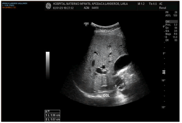
Figure 1: Ultrasound of admission where it is observed common bile duct of 1.9 cm with lithto inside with measurement of 1 cm.
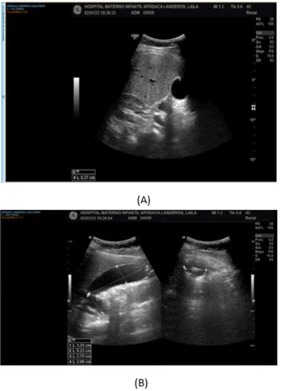
Figure 2: Admission ultrasound showing (A) gallbladder wall and measurements, (B) vesicle measurements as well as hyperechogenic images with posterior acoustic cobra referring to liths.
Case Presentation
This is a 14-year-old female patient who is admitted to the emergency department with jaundiced syndrome and abdominal pain, does not have a relevant personal pathological history for the current condition which begins with one month of evolution (November 25, 2022) with cramping abdominal pain in the right hypochondrium accompanied by jaundice and choluria, so she goes to a doctor where she is diagnosed with type A hepatitis, without seropositivity, receiving unspecified symptomatic treatment with temporary improvement, 25 days later acolia and abdominal pain in the right hypochondrium are added, so he goes again to a private doctor where ultrasound of the liver and bile ducts is performed where cholecystitis and dilation of the common bile duct are reported, as well as accentuation of abdominal pain, which is why he goes to the ISSEMyM Maternal and Child Hospital for assessment (Table 1).
Table 1. Chronicity of laboratory results at admission, beginning of antibiotic treatment and after surgical procedure, the marked difference in bilirubin levels and levels of leukocytes and neutrophils after both the establishment of antibiotics and surgical act is observed.
| Date / Labs | TP | INR | TPT | Leucos | NEUT | HB | HTO | PLQ | Urea | Creatinin A | BT | BD | BI | AlbuminA | FA | ALT | AST | PCR |
|---|---|---|---|---|---|---|---|---|---|---|---|---|---|---|---|---|---|---|
| ADMISSION | 13.4 | 1.1 | 29.5 | 8.44 | 73 | 10.5 | 31.8 | 389 | 21.4 | 0.51 | 17.6 | 13.4 | 4.2 | 3.5 | 685 | 192 | 180 | 56.4 |
| 04.01.23 | 13.3 | 1.1 | 29.9 | 12.35 | 78 | 15.4 | 44.8 | 404 | 12.8 | 0.52 | 16 | 12 | 4 | 3.2 | 529 | 175 | 205 | 58.7 |
| 05.01.23/ POST QX | 13.2 | 1.1 | 35.1 | 9.6 | 77 | 13.9 | 40.8 | 398 | 12.8 | 0.51 | 11.6 | 8.7 | 2.9 | 3.1 | 481 | 152 | 144 | 71.26 |
| 06.01.23 | 12.2 | 1.05 | 32.3 | 6.76 | 71 | 12.4 | 36.8 | 309 | 15 | 0.46 | 10.1 | 7.4 | 2.7 | 3 | 442 | 119 | 88 | |
| 09.01.23 | 12.8 | 1.1 | 31.6 | 6.1 | 69 | 12.4 | 34.5 | 318 | 25.7 | 0.69 | 9.3 | 6.7 | 2.6 | 3.8 | 364 | 118 | 131 | |
| 11.01.23 | 7.2 | 5.4 | 1.8 | |||||||||||||||
| 13.01.23/ ALTA | 8.31 | 66 | 12.1 | 34.6 | 270 | 5.4 | 4.1 | 1.3 | 3.9 | 309 | 94 | 88 | ||||||
| 17.01.23/AMBU | 11.3 | 1 | 33.7 | 9.13 | 12.3 | 36.3 | 390 | 34.2 | 0.54 | 4.6 | 3.5 | 1.1 | 4.5 | 289 | 107 | 90 |
He was admitted to the pediatric surgery service, presented respiratory deterioration, hemodynamic instability with amine requirement and shock data, so he was admitted to the intensive care unit with a diagnosis of choledocholithiasis + severe cholangitis.
Magnetic resonance cholangioresonance was performed as part of the study protocol and its transfer to a third level unit for ERCP was requested without obtaining a favorable response, derived from the state of septic shock (Figure 3). It was decided to perform laparotomy with bile duct exploration for bile duct drainage, with the following findings:
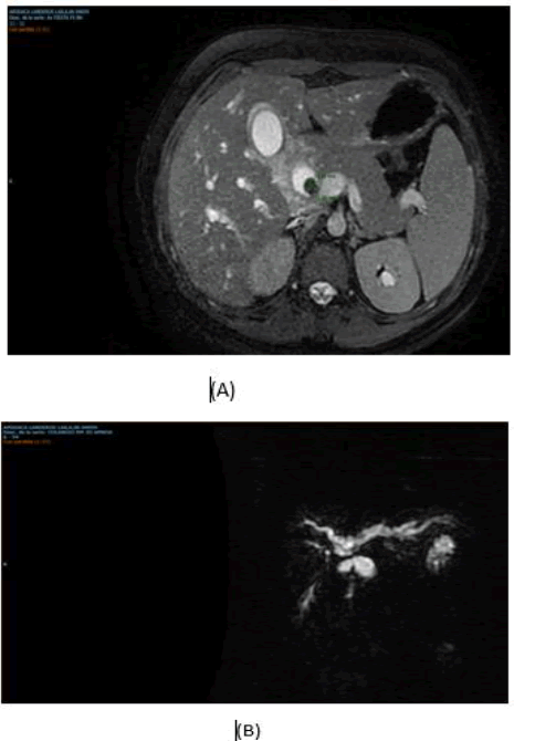
Figure 3: (A) Axial section cholangioresonance where the lithium is evident in the bile duct, (B) 3D reconstruction of cholangioresonance where amputation is observed at the confluence by liths at that level.
There is vesicle with multiple adhesions omentum background Zulkhe IIIIV, edematous and fibrous tissue with inflammatory reaction, thickened peritoneum, anatomy altered by inflammatory reaction, dilated common bile duct of approximately 2.8 cm longitudinal colectomy is performed with purulent bile outlet, drains and extracts 2 litos of approximately 1.5 cm each (choledocholithiasis of large elements). Sphincteroplasty with Baker's dilators with adequate passage is performed (Figure 4).
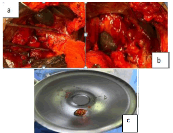
Figure 4: Surgical procedure, exploration of open bile duct: (a) The dilation of the common bile duct and the surrounding edematous tissue and inflammation are observed, (b) Longitudinal choledochotomy for exploration of the bile duct, (c) one of the extracted stones of 1.5 cm.
A T-probe is placed and transoperative cholangiography is performed with adequate passage to the duodenum and to the proximal biliary tract without filling defects, subtotal cholecystectomy is performed. Bleeding 250 cc (Figure 5).
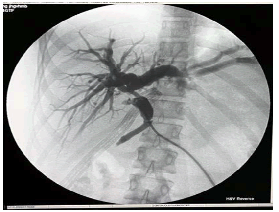
Figure 5: Transoperative cholangiography showing contrast medium without filling defect.
It presents adequate postoperative evolution, tolerating orally, serous drainage expenditure and scarce, with decreased bilirubin levels with low output through T-tube, control cholangiography is performed on the 4th day (Figure 6), which although even with dilation of the bile duct does not present filling defect and presents adequate passage to the duodenum, so drainage is removed and he is discharged from hospitalization, a T-probe is removed on day 28 with adequate laboratory controls and clinical evolution.
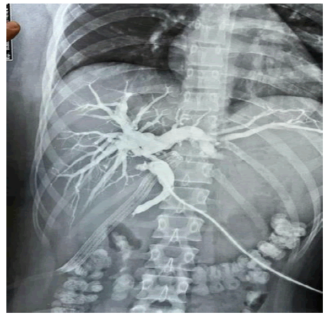
Figure 6: Transprobe cholangiography without filling defect and passage of contrast medium to the intestinal tract.
Choledocholithiasis defined as the presence of stones in the bile duct is considered a relatively rare entity in the pediatric population with a reported incidence of 0.15%-0.22% with increase during puberty, with the highest incidence reported at 11 years-15 years being more frequent in women, with a more prevalent risk factor such as obesity [1]. For the presumptive diagnosis of this entity, the criteria of the American Society for Gastrointestinal Endoscopy 2019 are used, which divide into high, intermediate or low probability of choledocholithiasis. According to the staging, has a positive predictive value of 85% and according to this, a therapeutic or diagnostic strategy to be followed is proposed [2-7] (Table 2).
Table 2. ASGE (American Society for Gastrointestinal Endoscopy) criteria. 2019.
| Probability | Predictors | Recommended Strategy |
|---|---|---|
| Alta | Evidence of common bile duct stone by Ultrasound/Imaging study Either Cholangitis Clinic either Total Bilirubin >4 mg/dl and Dilation of the Bile duct on Ultrasound/Imaging Study |
CPRE |
| Intermediate | Abnormal liver function tests either Patient older than 55 years either Common bile duct dilation on ultrasound/imaging study* |
Magnetic resonance cholangio/ endoscopic ultrasound/ Intraoperative cholangiography/Intraoperative ultrasound |
| Baja | No predictor present | Cholecystectomy with or without Intraoperative Cholangiography |
*Coledocus diameter>6mm
For this staging, a patient study protocol with laboratory studies that include coagulation times, blood biometrics, blood chemistry, liver function tests and ultrasound are necessary.
Likewise, diagnostic criteria for cholangitis must be used, for which the criteria of the Tokyo 2018 guidelines are used (Table 3).
Table 3. Cholangitis Diagnostic Criteria (Tokyo Guidelines 2018).
| Systemic inflammation | A1- Fever and/or chills A2-Biochemical Evidence of Inflammatory Response |
|---|---|
| Cholestasis | B1-Jaundice (Total bilirubin>2 mg/dl) B2- Abnormal Liver Function Tests |
| Image | C1- Bile duct dilation C2- Evidence of obstruction by image |
• Suspected Diagnosis: An item of A + An item of B or C ï?·Definitive Diagnosis: One item of A + One item of B + One item of C
Once the diagnosis of cholangitis is made, its severity is classified according to the Tokyo 2018 Guidelines and treatment is established (Table 4). Although there are certain therapeutic specifications regarding its severity, it is necessary to initiate support measures in general: fluid therapy, analgesia and the cornerstone of medical treatment is the antibiotic to which with respect to different guidelines should be started within the first hour mainly if it is associated with shock or a picture of severity Grade III [4-14].
Table 4. Cholangitis Diagnostic Criteria (Tokyo Guidelines 2018).
| Grade III (Severe) | Acute cholangitis that is associated with dysfunction of at least one of the following organs/systems | Cardiac: Hypotension with amine requirement Neurological: altered consciousness Respiratory: PaO2/FiO2 <300 Renal: Acute Kidney Injury Hepatic: INR >1.5 Hematologic: Platelet count< 100,000 |
|---|---|---|
| Grade II (Moderate) | Acute cholangitis in relation to any of the following conditions | Abnormal white blood cell count>12 or <4 Fever >75 years BT hyperbilirubinemia>5 mg/dl Hypoalbuminemia |
| Grade I ( Mild ) | Acute Cholangitis | No criteria for Grade II or III |
If classified as severe, it is recommended to follow the international guidelines of the campaign surviving sepsis for the treatment of septic shock in children, thus starting broad-spectrum antibiotic treatment preferably within the first hour of diagnosis, having lactate measurement and following an action protocol according to each institution. Following these guidelines, a combination of management can be made according to the management of sepsis and according to Tokyo 2018 taking into account the adjustment of dosage according to age and weight (Table 5) [5-14].
Table 5. Antibiotics for the management of cholecystitis and cholangitis.
| Grade/ Antibiotic | Based on Penicillin | Based on Cephalosporins | Based on Carbapenems | Based on Fluoroquinolone |
|---|---|---|---|---|
| Grade I | Piperacillin/Tazobactam | Cefazolina Cefuroxima Ceftriaxona Cefotaxima ± Metronidazol |
Ertapenem | Ciprofloxacin Levofloxacin moxifloxacin ± Metronidazole |
| Grade II | Piperacillin/Tazobactam | ceftriaxone cefotaxime Cefepime ± Metronidazole |
Ertapenem | Ciprofloxacin Levofloxacin moxifloxacin ± Metronidazole |
| Grade III | Piperacillin/Tazobactam | Cefepime Ceftazidime ± Metronidazole |
Imipenem/Cilastatin Meropenem ± Metronidazole | - |
| Associated with instrumentation or health care | Piperacillin/Tazobactam | Cefepime Ceftazidime ± Metronidazole |
Imipenem/Cilastatin Meropenem ± Metronidazole | - |
The study was approved by the Ethical Committee of the Faculty of Medicine, The specific treatment for cholangitis is based on drainage of the bile duct, which for some effect there are several approaches, which are: percutaneous liver puncture of the bile duct, exploration of the open or laparoscopic bile duct and Endoscopic Retrograde Cholangiopancreatography (ERCP) that although the recommendation is to perform associated procedure for the extraction of stones, If this is not possible, a stent is placed to achieve drainage of the bile duct.
Such procedures are recommended to be performed in a timely manner in order to reduce morbidity and mortality and hospital stay; The guidelines of the European Society of Gastrointestinal endoscopy recommend drainage of the bile duct in the first 12 hours of being a severe condition, within the first 48 hours-72 hours in moderate cases and selectively in mild cases, in such a way that the intervention to be performed has to be chosen according to the availability to achieve the treatment according to the previously commented time for better results and better survival [6,15].
In the case of severe cholangitis, higher rates of bleeding have been reported after sphincterotomy and balloon sweeps for the extraction of stones during ERCP, which is why the placement of stents alone is recommended, however given the relatively low percentage of complications 4%, it is important to individualize each case to have the planning to take, Since having bile stones of large elements (>1 cm) it is possible that the extraction of stones is not successful and the treatment is limited to drainage or planning for the subsequent extraction of said stones, which leads to the placement of endoprostheses and the replacement of them with previously planned times requiring multiple interventions, which is why it is recommended to perform drainage and resolution of choledocholithiasis in a while [7].
There are cases in which there are liths of difficult extraction that according to the literature is defined as liths of size >10 mm-20 mm, which are 2 or more liths of 1 cm, presence of distal lith, coexistence of diverticula, narrow distal prison or anatomical variants that condition sigmoid forms in the common bile duct where spyglass or bile duct exploration either open or laparoscopic has to be resorted to. There are comparative studies between these two procedures where greater success has been seen in resolving choledocholithiasis with bile duct exploration [8, 9]. However, the above in the context of hospital environment where there is material and human resources for such procedures, where there is no equipment to perform ERCP or Radio Intervention. We proceed to the exploration of the bile duct for the purpose of drainage of the route and control of the septic focus, in addition as mentioned above surgical intervention may be necessary for the resolution of choledocholithiasis pictures of difficult extraction with greater resolution success with these techniques [10].
In hospital units where there is no ERCP to be used urgently, and having a patient with severe cholangitis where drainage of the bile duct is urgently required, the exploration of the bile duct is recommended to achieve drainage of the same and with this the control of the septic focus.
According to the picture of choledocholithiasis, individualize the risk with respect to a procedure that resolves it, cholangitis and cholecystectomy, or only the drainage of the bile duct, this according to risk factors associated with the patient comorbidities and alterations due to the current condition, or manage to resolve in a single surgical time since it has less morbidity, shorter hospital stay and less need for material resources with a lower cost in care with a higher success rate as mentioned in the ASGE and Tokyo 2018 guidelines. Always following the points to achieve a safe cholecystectomy by performing an anterograde dissection "the bottom first" and / or perform subtotal cholecystectomy which is indicated in case where the dissection of the hepatocistic triangle is not safe and the biliary anatomical structures cannot be adequately identified, with an index of similar complications in terms of performing open or laparoscopy that for this purpose takes into account the preference and experience of the surgeon, with a recurrence of stones in 5% which justifies the procedure in whose context of the patient presented a reconstitutive cholecystectomy is performed. So it is concluded that it can be carried out safely and resolutely as it is presented in a previous case [11,13].
Acute cholecystitis, choledocholithiasis and cholangitis are pathologies that can coexist and according to their severity requires timely resolution having various procedures, times and surgical techniques to achieve this goal, the surgeon must be familiar with this range of therapeutic options since not always all the material resource is available and the time elapsed between diagnosis and treatment can be crucial for the evolution of the patient reason why you should have diagnostic suspicion and accurate therapeutic decisions.
In the pediatric population, the guidelines for biliary pathology focus mostly on congenital disorders such as biliary atresia, however there are no guidelines for the pathologies previously commented, which are also increasing with respect to their incidence, this article and the medical action are based on guidelines established for the adult population, Thus, it is concluded to extrapolate such guidelines and the development of new ones or to study the validity of existing ones in the pediatric population at different stages of growth and development.
The authors involved in this article declare that they have no conflict of interest.
Citation: : Cobos R.R.U., Rodas, E. Cholecystitis, Cholangitis and Choledocholithiasis: Presentation of Clinical Case in Pediatric Patients and Review of Guidelines. Surg.: Curr. Res., 2023, 13(6), 451.
Received: 09-May-2023, Manuscript No. scr-23-24014; Editor assigned: 11-May-2023, Pre QC No. scr-23-24014 (PQ); Reviewed: 23-May-2023, QC No. scr-23-24014 (Q; Revised: 28-May-2023, Manuscript No. scr-23-24014 (R); Published: 09-Jun-2023, DOI: 10.35248/2161-1076. 23.13.06.45
Copyright: �???�??�?�©2023 Cobos R.R.U.,This is an open-access article distributed under the terms of the Creative Commons Attribution License, which permits unrestricted use, distribution, and reproduction in any medium, provided the original author and source are credited.