Case Report - (2022) Volume 11, Issue 3
This paper presents a novel method of upper and lower jaw reconstruction using 3D-custom-made titanium implants with abutment-like projections. The implants were designed to rehabilitate the oral and facial shape, aesthetic, function, and occlusion. A 20- year-old male was diagnosed as having Gorlin syndrome. The patient suffered from having large bony defects, following ablative multiple keratocysts resection, of the maxilla and mandible. The resulting defects were reconstructed with 3D-custom-made titanium implants. The implants with abutment-like projections were simulated, printed, and fabricated with a selective milling method based on CT-scan data. There was no postoperative infection or foreign body reaction during the one-year follow-up period. To the best of our knowledge, this is the first report on the use of 3D-custom-made titanium implants with abutment-like projections, attempting to rehabilitate the occlusion and overcome the limitations of custom-made implants in treating large bony defects of the maxilla and mandible.
Custom-made implant • 3D Printing • Mandible reconstruction • Gorlin syndrome • Keratocyst
Reconstruction of the upper and lower jaws, and large bony defects, following ablative tumor surgery is one of the major challenges faced by the maxillofacial surgeon. The resulting defects of the mandible and maxilla, have a great impact on the aesthetics and oral functions such as mastication, swallowing, and speaking. Several reconstruction techniques have been used to achieve optimal functional and aesthetic results. When the defect is large, a micro vascular free flap has been the golden standard of mandibular and maxillary reconstruction [1-9]. Autogenous graft, however, has its disadvantages such as donor site morbidity, extended operation time, and potential graft failure due to tissue infection and necrosis. 3D-custom-made titanium implant has been successfully used for the reconstruction of facial bone defects including the mandible [7-9]. The advantage of a 3D-custom- made titanium implant is that it can be accurately designed according to the defect size and morphology. This will reduce the operative time and also recovers the original contour of the mandible and maxilla achieving optimal facial symmetry [1-6]. Also, the placement of 3D implants following ablative cancer surgery is an advantage, especially when the oncology treatment requires post-operative radiotherapy for disease control because it avoids the issue of secondary surgery in irradiated tissues. However, 3D-custom-made titanium implant has its limitations when treating large bony defects and rehabilitating the occlusion. This paper presents a novel method of upper and lower jaw reconstruction using 3D-custom-made titanium implants with abutment-like projections.The implants were designed to rehabilitate the oral and facial shape, aesthetic, function, and occlusion.
20 years old patient was referred to extract an impacted upper right wisdom tooth. There was no associated pain and/or swelling. Examination of the face showed frontal bossing, broad nasal bridge, hypertelorism, mandibular prognathism, and deformity of the lower extremities (Figure 1a, 1b).
Figure 1: 1a) Preoperative photo of the patient showing frontal bossing, broad nasal bridge, and hypertelorism of a 20-year-old patient with Gorlin syndrome, 1b) postoperative photo of the same patient following the reconstruction of the upper and lower jaw using 3D-custom-made implants achieving optimal appearance.
Virtual pre-surgical planning
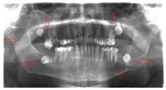
Computed Tomography Scan (CT-scan) and Orthopantomograph (OPG) revealed well-defined multiple, multilocular radiolucencies, with a sclerotic border involving the entire posterior maxilla and mandible (Figure 2).

Figure 2: Pre-operative Orthopantogram (OPG) shows multiple cystic lesions in the mandible and maxilla involving the maxillary sinus on either side with the displaced teeth (red arrows).
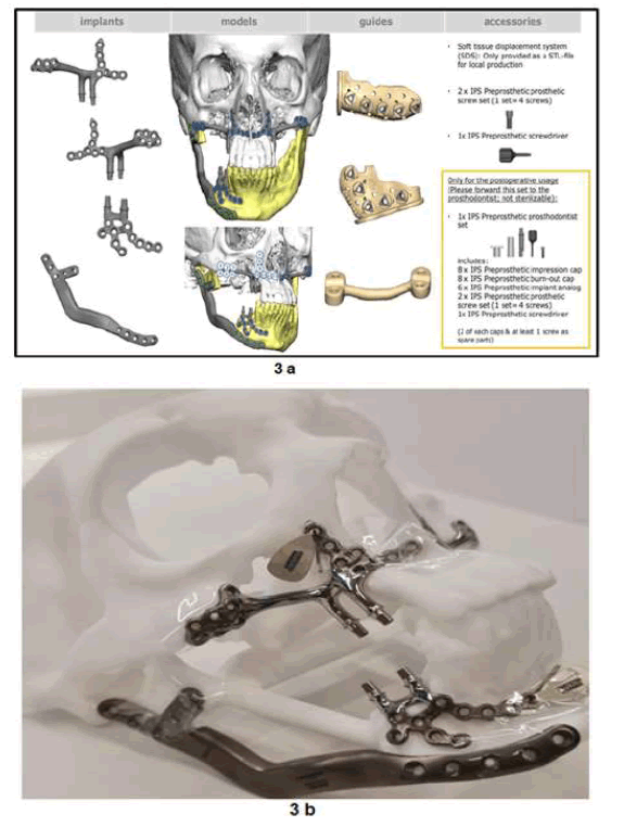
It was carried out based on CT-scan images (3D eXam, KaVo GmbH, Biberach, Germany) and virtual set-ups (3Matic Medical, Materialise, Leuven, Belgium). Resection surgical guides and titanium mandibular, maxillary implants were then fabricated using a 3D printer with a selective milling technique designed by KLS Martin (KLS Martin, Tuttlingen, Germany). The surfaces of the implants were polished. Multiple bicortical screw holes were designed on each end of the implant for fixation (Figures 3,4)

Figure 3: 3a) Diagram showing the virtual planning of the 3D-custommade implants together with drilling /surgical guides. The diagram also displays the prosthetic components for dental restoration, 3b) a Stereolithographic white model of the maxillofacial skeleton was also produced. Four implants were fabricated. Two mandibular implants with two abutment-like projections were used to restore the form, shape, and occlusion of the right mandible. The other two 3D-custom-made implants were used to reconstruct the right and left posterior maxilla with two abutment-like projections to restore the dentition
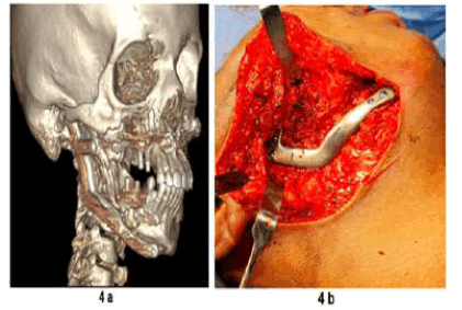
Surgery was carried out under general anesthesia. The right body of the mandible, including the ascending ramus, sparing the condylar head, was resected. Resection guides were well fitted on the mandible body according to the virtual plan. Subsequently, the titanium implant was inserted into the resected area and fixed using bicortical screws on the remaining mandible and subcondylar area (Figures 3, 4). To reduce the functional load, a second 3D-custom-made titanium mandibular implant was fabricated and fixed to the anterior mandible body (Figures 3, 4). For future dental rehabilitation, two abutment-like projections were printed into the second titanium mandibular implant with projections emerging above the soft tissue for future dental prosthesis. While the cystic lesion on the left side was fully enucleated, followed by curettage. The upper right and left posterior maxilla were fully enucleated/resected leaving large bony defects, sparing the anterior segment of the maxilla. The maxillary defects were reconstructed using 3D-custom-made implants (Figure 3, 4). The positioning of the maxillary implants was planned with the distal part of the implant fixed to the lateral cortical bone of the zygomatic complex. The proximal part of the implant was fixed to the following bone structures: alveolar bone of the anterior segment of the maxilla and lateral nasal wall (Figures 3, 4).

Figure 4: 4a) 3D image of the reconstructed mandible and maxilla using the 3D-custom-made implants, 4b) Intraoperative photo of the 3.0 mm 3Dcustom-made titanium implants fixed with 2.1 mm locking screws.
Reconstruction of large bony defects of the maxilla and mandible represents a challenge to the maxillofacial surgeon. Reviewing the literature several reports were published on the use of 3D printing techniques to reconstruct the mandible [1-9]. However, few studies were reported on the use of 3D-custom-made implants to reconstruct the maxilla. These mainly focused on the use of 3D sub-periosteal implants to reconstruct the atrophic maxilla. Rather than reconstructing large bony defects of the maxilla following ablative tumor surgery [6-10]. Mastication and pronunciation are important functions of the mandible and maxilla that can be restored with dental rehabilitation. The use of 3D-custom-made implants for occlusal rehabilitation is limited in that dental implants cannot be installed into the custom-made implant body itself during the fabrication process [1-6]. The exposure of the 3D-custom-made implant, following surgery to place dental implants, leads to infection of the implant and eventual failure. In this study, we used 3D-custom-made implants with two abutments like projections designed as part of the implant to rehabilitate the occlusion. To avoid the risk of infection the soft tissues surrounding the 3D-custom-made implants were closed with the abutment-like projection emerging through the soft tissue to avoid the second surgery and the increased risk of infection.
To the best of our knowledge, this is the first report on the use of 3Dcustom-made titanium implants with abutment-like projections, attempting to rehabilitate the occlusion and overcome the limitations of custom-made implants in treating large bony defects of the maxilla and mandible
Acknowledgement
Citation: Sukhun A. J. et al. A Novel Method To Reconstruct The Upper And Lower Jaws Using 3d-Custom-Made Titanium Implants. Reconstr Surg Anaplastol, 2022, 11(3), 001-003.
Received: 28-Jun-2022, Manuscript No. ACR-22-18902; Editor assigned: 30-Jun-2022, Pre QC No. ACR-22-18902 (PQ); Reviewed: 07-Jul-2022, QC No. ACR-22-18902 (Q); Revised: 15-Jul-2022, Manuscript No. ACR-22-18902 (R); Published: 28-Jul-2022, DOI: doi.10.37532/22.11.3.15-17
Copyright: ©2022 Sukhun A. J. This is an open-access article distributed under the terms of the Creative Commons Attribution License CC-BY, which permits unrestricted use, distribution, and reproduction in any medium, provided the original author and source are credited.