Research Article - (2016) Volume 5, Issue 2
Kavir-48, a protein with a molecular weight of 44 KDa, isolated from an extremophile Streptomyces was investigated for anti-proliferative activity on 10 human cell lines.
5000 cells were plated in 96 well plates. After 24 h. Kavir-48 was added in five dose levels (0.01-100 μg/ml). After 4 days of continuous exposure, the cellular DNA content and morphology was determined, using propidium iodide staining, the resulting fluorescence correlates with a number of leased cells.
A selective growth inhibition (Mean IC 70) observed for the large cell Lung cancer cell line and mammary cell line. The antitumor activity as well as tolerability of product was evaluated in in vivo testing on lung cancer cells, (Xenograft in nude mice) since there might be a selective antitumor effect after 21 days of administration. Neither lethality nor body weight loss was observed in all group of tested animal, and in the large cell lung cancer model, a clear anti tumor effect observed after only five days of administration. Amino acid sequencing of protein suggest that the compound consist of a fusion proteins NISOD and a Tat protein combination.
SDO are generally classified according to the metal species as well as, Nickel containing SOD (NiSOD), of Streptomyces spp.
Expression of the enzymatic activity of the transduced Tat-SOD fusion proteins is essential for therapeutic application; for several reasons, such as the size and instability of the SOD enzyme these attempts to develop the natural enzyme for clinical use have been largely unsuccessful.
The differential effect of Kavir-48 against distinct tumor cell lines may be due to the fact that this protein is a Fusion of Tat and NiSOD protein and then it is possible that the binding of the protein to a cell type specific receptor may trigger apoptotic signal.
Keywords: Streptomyses; Anticancer; Apoptosis; Superoxide dismutase; Tat- protein; Xenograft; Kavir-48; Oxidative stress
A wide variety of physiological and pathological stimuli can initiate apoptosis. They act via receptor mechanisms, through biochemical agents, or cause DNA and cell membrane damage, followed by degradation of the engulfed cell DNA. Apoptotic cells undergo a series of change including: Shrink in size, fragmentation of DNA, creating a vacuolar nucleus, cytoplasm break into smaller pieces called apoptotic bodies.
Living organisms have been a major source of new biologically active molecules for the pharmaceutical, animal health and agrochemical industries for much of this century.
After the discovery of penicillin in 1929, screening of secondary metabolites of Microorganisms as a natural source of potential drug candidates were explored.
Among the well characterized pharmaceutically relevant microorganisms, Actinomycetes and Streptomyces remain the major sources of novel, therapeutically useful natural products [1,2].
They produce over two thirds of the clinically useful antibiotics of natural origin.
Streptomyces is a genus of Gram-positive bacteria that grows in various environments. The most interesting property of Streptomyces is the ability to produce bioactive secondary metabolites, such as antifungals, antivirals, antitumorals, anti-hypertensives, immunosuppressants and especially antibiotics. The production of most antibiotics is species specific, and these secondary metabolites are important for Streptomyces species in order to compete with other microorganisms in the environment.
They are classified by the interaction of bioactive molecules, targeting essential cellular functions, the fundamental principle to inhibit cell growth. This is a complex process that starts with the physical interaction of the molecule and its specific targets and involves biochemical, molecular, and structural changes, acting on multiple cellular targets such as DNA replication.
Programmed cell death or apoptosis is an integral part of differentiation program. Many of cancer cells fail to do so [3]. Most of the drugs currently used in cancer treatment either damage DNA or inhibit DNA replication. In this case the failure to undergo apoptosis contributes to the resistance of cancer cells to irradiation and of many chemotherapeutic drugs, which act by damaging DNA. The possibility exists of developing drugs that interact with components of the cell cycle machinery, including apoptosis.
In the case of screening for new anticancer agents we have isolated a new potent anticancer compound that might induce apoptosis of temporal cells. In this communication we report in vitro and in vivo effects of the anticancer compound, Kavir-48 and the partially identification of their chemical structure,
Human tumor Xenografts are being considered as the most relevant models for anticancer drug development. In addition the Xenografted tumor models resemble very closely the original tumors regarding histology and a high grade of correct productiveness of the Xenograft models for profiling anti tumor test compounds for subsequent clinical studies.
Strains
Kavir-48 is a protein of 44KDa isolated in the course of a screening program for new anticancer agents from a microbial strain isolated from Central Desert of IRAN, taxonomically assigned to the genus Streptomyces, identified as Streptomyces anulatus and deposit in Persian Type Culture Collection: (PTCC-1660).
Test compounds
The sample was activated in ISP2 medium (Difco™ Dehydrated Culture Media: ISP Medium 2) enriched with 5% of NaCl. Fermentation of isolated Streptomyces was run in rotary incubator shaker in a medium consisting: Yeast extracts 0.3g/l; Beef extract 0.3 g/l; Tryptose 0.8 g/l FeSO4.7H2O, Glucose 3.5 g/l. PH was adjusted to 7.2, for 120 h at 28ºC and 150 rpm. Supernatant was harvested by centrifugation, in 7000 g, (Beckman model J2-21) and filtrated supernatant was use for anticancer screening.
E. coli ATCC 33312, the lysogenic indicator strain was developed for screening of anticancer sample by prophage induction assay. Cytotoxicity against Murine Leukemia P388 cell line was measured by the percentage of cell death and cancerous cells without product use as positive control.
In vitro assay
P388 Murine leukemia cell line were maintained at 37ºC in 90% DMEM containing HAT (Thymidin 0.14 μ m, Aminoterin 40 μ m and hypoxanthin 0.1 μ m) and 10% fetal calf serum under a humidified atmosphere of 5% CO2, 95% air. Cell were plated in 96 well microplate at a density of 5000 cells per well with 100 μl media and 10 μl, 25 μl, 50 μl and 100 μl of product.
The plate was incubated in the CO2 incubator at 37ºC for 48 h. Every 12 h the well prepared smear was stained with Gimsa. The sample present apoptosis on P388 cell line was examined in vitro in a panel of 10 human tumor cell lines (Table 1).
| Tumor Type | Cell lines | Histology | Doubling time | Tumor formation in vivo |
|---|---|---|---|---|
| Colon | CCHT29 | Pdadeno Ca. | 23 | yes |
| Gastric | GXF 25L | Pdadeno Ca. | 32 | yes |
| Lung NSCI | H 460 | Large Cell | 18 | yes |
| Lung NSCI | LXFA 629 L | Adeno. Ca. | 31 | yes |
| Lung NSCI | LXFL 529 L | Large cell | 25 | yes |
| Lung NSCI | LXFE 66NL | Squamous cell Ca. | 41 | yes |
| Mammary | MCF-7 | Adeno Ca. | 30 | yes |
| Melanoma | MEXF276 NL | Amelanotic | 33 | yes |
| Renal | RXF 994 L | Carcinoma | 30 | yes |
| Uterus | UXF 1138 L | Carcino-Sarcoma | 31 | yes |
Table 1: Human tumor cell lines used for the in-vitro testing of kavir-48.
In this assay, cell is exposed continuously to the drug for four days corresponding to four doubling times of the tumor cells. After the incubation period, vital cells are quantified by means of the propidium iodide assay in comparison to the well-known sulforhodamin B staining of cellular protein.
In vivo assay
The antitumor activity as well as tolerability of Kavir-48 was evaluated in vivo in the large cell cancer models LXFl 529 and LXFL H460.
For Xenograft of LXFL 529 and LXFL eight to ten weeks old male and for H460 Xenogradt, eight to ten weeks female nude mice of NMRI genetic background were used. Mice were randomly assigned to treatment groups and the control group (6 mice in each group). Tumor fragments of about 20 mg were implanted subcutaneously in both flanks of athymic nude mice.
Treatment was started after an induction time of 12 days, when the median tumor volume ranged for 89 to 122 mm2 for Xenograft LXFL 529 and induction time of 10 days, when the median tumor volume ranged from 149 to 164 mm2 for Xenograft LXFL H460.
Kavir -48 was administrated i.p. in tumor bearing nude mice at doses of 150 mg/kg/day and 300 mg/kg/day. On 5 (in the case of LXFL 529) or 10 consecutive days with an intermediate break of two days after 5 days (in the case of LXFL H460). To determine the maximum tolerated dose (MTD) in an orientating approach with two animals per dose group, nude mice without tumors were treated with Kavir-48 and the body weight course and lethality was recorded.
First Kavir-48 was administrated at dose 3, 10 and 30 mg/kg twice a week. On day 1, 5 and 9, and animals were observed for 21 days. After 21 days, neither lethality nor body weight loss was observed. The mice of the control group survived until the end of the experiment and gained body weight to a relative median value of 116.7% of the initial body weight. The doses were then increased to 30, 100 and 300 mg/kg.
Mortality checks were conducted once daily. Body weight of the mice was determined two times a week i.e. at the same day when the tumors were measured.
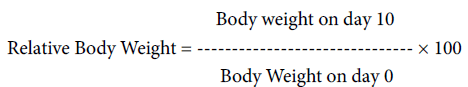
The body weight of the treatment group was expressed at the relative median body weight of all animals of the group. The relative body weight of individual animal on day 10 was calculated according to the formula:
The tumor volume was determined by two dimensional measurement with a caliper at the day of randomization (day 0) and then twice weekly. The tumor volume was calculated according to the formula:
Tumor volume = (a × b2) × 0.5
Where a and b represent two perpendicular tumor diameters: a=length (larger diameter) and b=width (smaller diameter).
The tumor reached mean diameters of approximately 18 mm in the mice of control group. The anti tumor effect was evaluated following maximal tumor inhibition versus the control group. Median Relative Tumor Volume (RTV) values were used for drawing growth curves and treatment evaluation.
Optimal growth inhibition at a particular day within the experimental period was calculated from the median RTV values of the test versus control groups multiplied by 100% (T/C% value).

Figure 1: Murine leukemia cells: a– control. b- 16 h. after treatment, cell is shrunken and develops blebs. Membrane integrity is not lost until late. c- 24 h. after treatment, nuclear chromatin undergoes condensation and fragmentation. d-36 h. after treatment, the cytoplasm becomes divided to form apoptotic bodies containing organelles and/or nuclear debris.
Kavir-48 showed promising activity, that indicate the compound might induce apoptosis in P388 cell line, (Figure 1) in vitro investigations of the protein using 10 different permanent human tumor cell lines were carried out (Table 1).
A selective growth inhibition (IC 70 mean IC 70/3), observed for the large cell Lung cancer cell lines, LXFL529 and H 460 was observed. The rest of the cell lines of panel reacted insensitive against treatment with Kavir-48.
The antitumor potency of Kavir-48 is on the basis of the molar mean IC 70, lies in the range of that of Mytomycin in a comparable tumor test panel. Cisplatin (0.84 μm), 5-Fluo-Uracil (5 μm) and Etoposide (2.17 μm).are less potent than Kavir-48, whereas Doxorubicin (0.17 μm), Topotecan (0.15 μm), Paciltaxel (0.11 μm) and Vindesine (0,007 μm) show higher antitumor potency than Kavir-48.
To determine the maximum tolerated dose (MTD) in an orientating approach with two animals per dose group, nude mice without tumors were treated with Kavir-48 and the body weight course and lethality was recorded.
First Kavir-48 was administrated at dose 3, 10 and 30 mg/kg twice a week. On day 1, 5 and 9 and animals were observed for 21 days. After 21 days, neither lethality nor body weight loss was observed. The mice of the control group survived until the end of the experiment and gained body weight to a relative median value of 116.7% of the initial body weight. The doses were then increased to 30, 150 and 300 mg/kg.
Was given twice a week on day 0, 4 and 8, animal were observed for 28 days. After the therapy and 14 days of observation, there was a slight body weight loss in the 300 mg/kg group to 93.8% of the initial body weight.
The schedule was the changed to a daily administration on day 14 to 18 to achieve a higher plasma level of compound. In the further course of the experiment one mouse died in the 300 mg/kg group on day 18, and one mouse died in the 150 mg/kg on day 25. The remnant animal in 300 mg/kg group lost body weight to 66.8% of the initial body weight on day 21, but recovered until day 28 to 93.8% of the initial body weight.
In the 150 mg/kg group, the median body course showed a loss until day 21 with 93.5% of the initial body weight and a recovery to 103.8% on day 28. Kavir -48 was in general well tolerated. The mice of the control group gained weight with a median value of 126.6% of the initial body weight, the group treated with Kavir-48 at 300 mg/kg/day also gained to 93.8 % of initial body weight.
Kavir-48 was tested in two human xenograft of the large cell lung cancer model LXFL 529 and LXFL H460.
To determine the maximum tolerated dose (MTD) in an orientating approach with two animals per dose group, nude mice without tumors were treated with Kavir-48 and the body weight course and lethality was recorded.
First Kavir-48 was administrated at 150 and 300 mg/kg twice a week. After 21 days, neither lethality nor body weight loss was observed. The mice of the control group survived until the end of the experiment and gained body weight to a relative median value of 116.7% of the initial body weight.
The mice of the control group gained weight with a median value of 126.6% of the initial body weight, the group treated with Kavir-48 at 300 mg/kg/day also gained to 93.8 % of initial body weight.
In the large cell lung cancer model LXFL 529, a clear anti tumor effect could be observed after only five days of administration (Figures 2 and 3).

Figure 2: Anti tumor activity of Kavir 48 on LXFL 529 treated mouse and control.

Figure 3: Relative median value of body weight, in treated animal and control.
As show in Figure 2, in LXFL 529 xenograft animals, Kavir-48 given at 300 mg/kg/day, effected a reduction of tumor volume to median value of 112.6% of the initial tumor volume, compared to 170.1% of the control group, resulting in T/C value of 72%. An optimum effect was observed after 3 days, when the relative tumor volume in the group treated with Kavir-48 at 300 mg/kg/day was 413.6% compared with 512.0% of the control, resulting in a T/C value of 68.8% in treated animals.
As show in Figure 3, there was only a marginal change in doubling times when animal carrying the H460 xenograft that were treated with Kavir-48. Whereas the anti tumor activity in the large cell lung cancer LXFL H460 was only marginally pronounced.
Amino acid sequencing of Purified compound reviled that the product consist of an NiSOD fusion protein (Figures 4-6).

Figure 4: Anti tumor activity of Kavir 48 on LXFL –H460 treated animal and control.

Figure 5: Relative median value of body weight, in LXFL–H460 treated animal and control.

Figure 6: Protein structure.
SDO are generally classified according to the metal species which acts as redox-active center to catalyze the dismutation reaction. Until recently, different metal species have been found; copper and Zinc containing SOD (Cu, Zn SOD), Manganese containing SOD (MnSOD), Iron containing SOD (FeSOD) and Nickel containing SOD (NiSOD), of Streptomyces spp.
NiSOd was suggested to represent a novel class of superoxide dismutase on its own since no amino acid sequence homology is found to enzyme s of the already existing classes.
Streptomyces NiSOD is located in the cytoplasm. This NiSOD might therefore be universally distributed in the genus Streptomyces [4].
Nickel SOD were composed of four identical subunits of 13.4 KDa, were stable at pH 4.0-8.0 and up to 70ºC. The absorption of enzyme exihibiting was maxima at 276 and 378 nm, and a broad peak at 531 nm.
The molecular mass of SOD was 13.4 KDa. Molecular mass of dimeric form NiSOD determined by SDS-page was 23.6 KDa. Even most SOD in bacteria is usually dimeric form, although tetrameric forms were reported in FeSOD and MnSOD from higher organisms.
Heterologous proteins such as the human tissue plasminogene activator, murine scFv and Fab antibody fragments were found to require the Tat pathway for the export of functional protein to the periplasm. Activity of this newly discovered pathway was found to be independent of ATP as an energy source and relied solely on the proton gradient present in the membrane.
Bacteria need to transport protein across their membrane for constant interaction with their extracellular milieu. Machinery for exporting folded proteins is available via the “Twin Arginine Translocase” (Tat) pathway.
The direct intracellular delivery of proteins has been difficult to achieve, due of bioavailibity barrier of the plasma membrane. During the past decade, a variety of peptides called protein transduction domains (PTDs) or cell penetrating peptides (CPPs), have been characterized for their ability to translocate into live cells. Recently, poly arginine exhibit even greater efficiency in term of delivery of several peptides and protein.
However recent report showed that some proteins could bypass the molecular cut-off imposed by the cell membrane, opening new possibilities in the field of molecular therapy.
Small region of protein and synthetic peptides called protein transduction domains (PTDs) can traverse biological membrane efficiently in a process termed PTD, were recently identified in HSV-1 (VP22). HIV-1(Tat). PTDs can traverse biological membranes efficiently in a process termed protein transduction: delivering their cargo into the cell [5,6].
The Tat pathway in Streptomyces species seems to be the major route of protein translocation, as the majority of secreted proteins are translocated via this system [7-9].
It was originally assumed that the Tat pathway was dedicated primarily to the export of proteins that bind complex redox-active cofactors, which by necessity are inserted into the preprotein in the cytoplasmic compartment [10].
The third general protein transport system found in Streptomyces, the Tat pathway, differs from Sec in that it transports prefolded proteins across the cytoplasmic membrane. It was originally assumed that the Tat pathway was dedicated primarily to the export of proteins that bind complex redox-active cofactors, which by necessity are inserted into the preprotein in the cytoplasmic compartment [11]. The Tat pathway in Streptomyces species seems to be the major route of protein translocation, as the majority of secreted proteins are translocated via this system. However, Streptomyces species and some other prokaryotes, including certain halophilic archaea, appear to export large numbers of non-cofactor-containing proteins by this pathway [6,12]. The animal experiments shown that Tat – apoptosis protein could inhibit the growth of tumor and the purified Tatapoptosis protein produced by E. coli has an antitumor effect by pharmacological assay [13].
It was originally assumed that the Tat pathway was dedicated primarily to the export of proteins that bind complex redox-active cofactors, which by necessity are inserted into the preprotein in the cytoplasmic compartment [9].
Tat-Cu, Zn- Superoxide dismutase (Tat-SOD), a major antioxidant enzyme, can be directly transduced into mammalian cells and skin [6].
Superoxide dismutase is an enzyme that is important to cells for converting potentially damaging free radicals into hydrogen peroxide and oxygen.
Recent reports suggest that elevated MnSOD activity may result in cell cycle arrest, apoptosis, or senescence [14,15].
These reports have been demonstrated that oxidants may play a role not only on inducer but also as mediator of the apoptosis process, and new therapeutic strategies that take advantage of increased reactive oxygen species or inhibition of endogenous antioxidant defence, producing a state of oxidative stress selectively in cancer cells have gained importance.
Alkylating agents and reactive isotopes are well known to kill cancer cells by the mechanism of oxidative stress [16].
It has been proposed that this phenomenon is due to the derivation of cancer cells from stem cells that have innately lower levels of SOD [12]. Furthermore, the Mn-SOD content could reflect the P53 mediated apoptosis through a bcl-2-independent pathway [17].
Alkylating agents and reactive isotopes are well known to kill cancer cells by the mechanism of oxidative stress [18].
SDO are generally classified according to the metal species which acts as redox-active center to catalyze the dismutation reaction [12,19]. Until recently, different metal species have been found; copper and Zinc containing SOD (Cu, Zn SOD),
Kavir-48 is a 44KDa protein isolated from a strain of genus Streptomyces. Studies show that this protein may induce apoptosis in cancer cell lines and induce cytotoxicity especially in tumor cell lines of large lung cancer.
One likely explanation for these results may be the protein structure, active principle of this protein itself, a distinct fragment or metabolism of protein of Kavir-48.
Kavir–48 is safe, after 21 days of administration, neither lethality nor body weight loss was observed in all group of tested animals. The dose of 300 mg/kg/day was in general well tolerated.
In the large cell lung cancer model LXFL 529, a clear anti tumor effect could be observed after only five days of administration.
The potential for the therapeutic use of proteins was limited by the impermeable nature of the cell membrane to such macromolecules.
Tat-Cu, Zn- Superoxide dismutase (Tat-SOD), a major antioxidant enzyme, can be directly transduced into mammalian cells and skin [20,21].
Recent reports suggest that elevated MnSOD activity may result in cell cycle arrest, apoptosis, or senescence [13,15].
Expression of the enzymatic activity of the transduced Tat-SOD fusion proteins is essential for any therapeutic application. For several reasons, such as the size and instability of the SOD enzyme, these attempts to develop the natural enzyme for clinical use have been largely unsuccessful.
These reports have been demonstrated that oxidants may play a role not only on inducer but also as mediocre of the apoptosis process. Alkylating agents and reactive isotopes are well known to kill cancer cells by the mechanism of oxidative stress [22]. One likely explanation for these results may be the protein structure, active principle of this protein itself, a distinct fragment or metabolism of Kavir-48 protein.
Anthracyclines, such as Epirubicin, are often used in treatment of malignant mesothelioma. These compounds produce oxidant through redox-cycling reaction and induce apoptosis [17].
However, there is a concern for selective production of oxidative stress in cancer cell, without exhibiting significant cytotoxicity in the normal cell.
The differential effect of Kavir-48 against distinct tumor cell lines may be due to the fact that this protein is a Fusion of Tat and NiSOD protein and then it is possible that the binding of the protein to a cell type specific receptor may trigger apoptotic signal.
In the current study, we have recognized a system of Tat- Nickel- SOD, elicits the outset of apoptosis presumably through its pro-oxidant property generating H2O2.
No toxicity or evidence of other unwanted pharmacological effects of compound was noted at either level supplementation.
Furthermore, the Ic50 values that are comparable to those of the clinically used drug Cis Platin and the relatively unique structure found in this compound may serve to provide a lead structure for the development further compounds for cancer chemotherapeutic potential.
The Center for International Scientific Studies and Collaboration of Ministry of Science, Research and Technology of Iran is gratefully acknowledged for providing financial support to carry out this research work.
