Research Article - (2023) Volume 12, Issue 1
Introduction: The most common surgical technique in implant-based breast reconstruction is the two-stage approach, positioning the expander in the submuscular plane. In our series, the inferolateral aspect of the implant was covered by the Serratus Anterior Muscle (SAM) or Serratus Anterior Fascia (SAF), preserving the muscle. The aim of this retrospective cohort study is to compare the two techniques in terms of morbidity, complications, and reconstructive outcome.
Materials And Methods: 196 women underwent mastectomy and immediate breast reconstruction for breast cancer referring to our center from 2017 to 2018 were enrolled in the study. Patients were divided into two groups, according to the technique used to cover the inferolateral aspect of the implant: 103 women were in the SAM group, and 83 in the SAF group.
Results: In SAM group, 3 women had postoperative bleeding, requiring surgery. In the SAF group, only 1 woman needed a surgical revision. Comparing the two techniques, length of hospitalization stays, and post-operative pain were statistically significantly lower in the SAF group than in the SAM group (p<0.05).
Conclusions: SAF flap could be considered a safe and effective technique compared to the use of SAM to cover the inferolateral part of the implant, allowing to achieve a good reconstructive outcome and a good quality of life. However, larger studies are needed to confirm our results.
Level III: Evidence obtained from well-designed cohort or case-control analytic studies, preferably from more than one center or research group.
Brest reconstruction • Brest reconstruction • Breast Surgery • Breast Cancer • Serratus Anterior Fascia Flap
Oncologic and reconstructive advancements in the management of patients with breast cancer have changed over the past century. Nowadays breast reconstruction is increasingly present in the treatment of breast cancer and many options can be evaluated to achieve are con-structed breast as natural as possible for shape, size, and feel [1]. Together with the diffusion of breast reconstructive techniques, several conservative mastectomy approaches have been developed (skinsparing, nipple-areola complex (NAC)-sparing and skin-reducing mastectomies) to allow an immediate reconstruction with better aesthetic results [2]. In general, reconstructive techniques can be divided into implant-based and autologous tissue flap breast reconstruction [3]. Implant-Based Breast Reconstruction (IBR) can be performed at the time of the mastectomy (immediate) or during a subsequent operation (delayed) [4-5]. Two different approaches could be used performing immediate breast reconstruction: in the one-stage technique the definitive implant is immediately placed; in the two-stage technique the expander placement is followed by final prosthesis insertion [6-7]. As described by Cordeiro et al, in two-stage implant-based breast reconstruction fundamental concepts are the musculofascial pocket and the inferolateral site [6]. The traditional approach consists of creating a pocket for the expander or the implant to optimize the muscular coverage. Its inferolateral site plays a central role, maintaining the implant in the correct position and recreating the lateral part of the inframammary fold [8]. Several options are available to obtain an optimal coverage of the implant inferolateral aspect including some muscle flaps, as the Serratus Anterior Muscle (SAM) and its fascia and allograft materials [9-10]. However, raising the serratus muscle off the ribs is painful and not ideally suited for define the inferolateral pole of the reconstructed breast. For this reason, this surgical technique has been improved to create a musculofascial pocket including only a portion of the serratus muscle and its overlying fascia to minimize morbidity. This approach provides two benefits: structural support to the expander, and a well-vascularized tissue for good expansion, preventing loss of the reconstruction caused by mastectomy flap necrosis. However, the muscular flap is often associated with donor site morbidity [6]. Recently, the use of Serratus Anterior Fascia (SAF) has been described to recreate the lateral mammary fold, to prevent the lateralization of the expander, avoiding muscular detachment, and decreasing post-operative morbidity [11].
The aim of this study is to compare the use of SAM and SAF to cover the inferolateral aspect of the implant in terms of morbidity, complications, and reconstructiveoutcomes.
This is an observational retrospective cohort study comparing morbidity, complications, and reconstructive outcomes in expanderbased breast reconstruction using Serratus Anterior Muscle (SAM) or Serratus Anterior Fascia (SAF) for the coverage of the inferolateral pole of the pocket. The institutional ethical board approved the study, and the informed consent was obtained under the institutional review board policies of hospital administration.
Consecutive women referrals to our center between January 2017 and December 2018 were enrolled in the study. Inclusion criteria were unilateral or bilateral breast cancer, needing unilateral or bilateral mastectomy, and immediate two-stage implant-based reconstruction. Exclusion criteria were inflammatory breast cancer, skin infiltration or fixation, and previous radiation. All the patients included in the study underwent psycho-oncological consultation before undergoing surgery. Age, number of drainages, the time when drainages were removed, intraoperative saline fill volume, post-operative complications and pain, and reconstructive outcome were collected for each patient. Complications were assessed evaluating major complications, needing a surgical revision, and minor complications, requiring only medical treatment, and they were classified using Dindo Clavien classification, until one month after surgery [12,13]. Post-operative pain was measured with a Visual Analogue Scale (VAS) scoring pain from 1 (no pain) to 10 (strong pain) on the 5th post-operative day. The reconstructive outcome was evaluated considering women’ satisfaction grade one month after surgery. Surgery was performed by 2 general surgeons, and one plastic surgeon (one for the SAM and one for the SAF technique). Patients underwent NAC-Sparing (NSM), Skin-Sparing (SSM), or skin-reducing mastectomy. In NSM, an incision of 6 cm to 8 cm was performed in the lateral portion of the breast. In SSM, the incision was angled upward toward the axilla to make the upper and lower skin flaps of similar length. Skin-reducing mastectomies were performed according to the technique described by Nava and colleagues [14]. A separate axillary incision was performed for the sentinel lymph node biopsy or Axillary Lymph Node Dissection (ALDN) when needed. Intraoperative histologic analysis of sections from sub-nipple-areola complex tissue was routinely performed in NSM. After mastectomy, the fascia of the pectoralis major and serratus anterior muscle was cautiously preserved while performing the mastectomy. The implant pocket was then created through an incision along the lateral border of the pectoralis major muscle and progressive dissection beneath the pectoralis major itself, continuing the inferior part of the dissection beneath the serratus anterior. Then, the sternal attachments of the pectoralis major were dissected from the second intercostal space to the inferior edge of the pocket. The lower attachments of the pectoralis major and the serratus anterior muscle were dissected at the level of the contralateral inframammary fold. The pocket extended into the deep fascial layer, avoiding direct continuity with the mastectomy site, in order to allow the best expansion of the lower breast pole. A partially inflated (around 20% final volume) expander was then inserted into the submuscular pocket. In the SAM group, the inferolateral aspect of the implant pocket was created harvesting the serratus anterior muscle fibers. In the SAF group, only the serratus anterior fascia was used, after having carefully separated it from the serratus muscle fibers. Two negative suction drains were always placed on each side, the first in the submuscular pocket and the second in the subcutaneous plane. Discharged criteria were: good pain control, absence of surgical site complications, absence of major complications, and serous fluid drainage. Drainage removal was considered when the daily volume was lower than 30 ml. The inflation of the expander started at least 15 days after the surgical procedure, then filling the expander every one or two weeks, according to the patient’s compliance, until the complete inflation volume was reached. The final implant insertion was performed by another team of plastic surgeons.The follow-up was continued till the reach of the ideal filling volume.
Statistical analysis
Quantitative variables were analyzed using the Wilcoxon and MannWhitney tests to detect differences between the two groups. Categorical variables were analyzed using the Chi-square test and Fisher’s exact test. A p-value of 0.05 or less was considered statistically significant. R software (version 4.0.3, R Foundation for Statistical Computing) was used for statistical analysis.
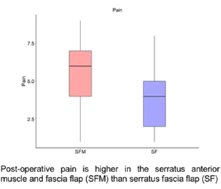
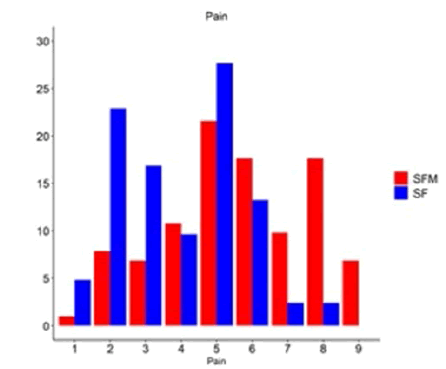
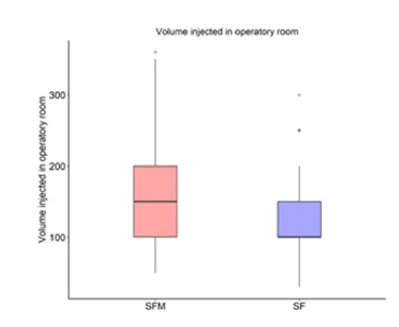
186 women were included in the study. All patients underwent a unilateral or bilateral mastectomy and two-stage implant-based breast reconstruction. In women undergoing surgery in 2017, the SAM was used to cover the inferolateral pole of the expander. Instead, in women undergoing surgery in 2018, the SAF was used. Results are shown in Table 1 and Table 2.The SAM group was made up by 103 patients. All patients underwent immediate positioning of a tissue expander with a mean filling of 156 ml (±63 ml). In this group, the mean Length Of Hospital Stays (LOS) was 2,5 days (±0.6day). The mean pain on the 5th post-operative day was 5 (± 2), 9 patients described 9 as pain value. We had a total complication rate of 31%: 10% of patients (10.3) had a seroma, 9.3% a hematoma (9), we did not have any abscess, 9.3% (9) had superficial skin necrosis. Using Dindo Clavien Classification for the complications rate, we divided it into minor (< 3) and major (> or = 3). Only 3 patients had a Dindo Clavien 3b complication needing a surgical revision (postoperative bleeding). In the SAF group, 83 women were enrolled. All the patients underwent immediate positioning of a tissue expander with a mean filling of 123 ml (±50 ml). The mean LOS was 2,2 days (±0.4day); and the mean pain on the 5th postoperative day was 3.9 (±1.7), with only one patient that described a pain value of 8 as pain value. The complication rate for this group was 14.4% (15): 3.3% of patie-nts (4) had a seroma, 2,5% (3) a hematoma, 5.8% (7) had superficial skin necrosis. Only one patient developed a Dindo Clavien 3b complication that needed a surgical treatment (postoperative bleeding). The statistical analysis between the SAF and SAM groups does not confirm a statistical difference concerning the total complication rate (p >0.1). The SAF group had a lower postoperative pain on the 5th day (Figure 1 and Figure 2) and a lower LOS than the SAM group (p <0.01 and p=0.01, respectively). Moreover, there was a statistically significant difference between the two groups in terms of volume injected in the operatory room (p<0.01) (Figure 3). This result is related to the fact that 2 different plastic surgeons performed the reconstructive phase, one for the SAM group and one for the SAF.
Table 1. Complications rate in the two groups.
| Type of complication | SFM Group (103) | SF Group (83) | p Value |
|---|---|---|---|
| Number of patients (%) | |||
| Minor Complication | |||
| Seroma | 10 (10.3%) | 4 (3.3%) | >0.1 |
| Hematoma | 9 (9,3%) | 3 (2.5%) | >0.1 |
| Superficial necrosis | 9 (9.3%) | 7 (5.8%) | >0.1 |
| Major Complication | |||
| Infection | 0 | 0 | |
| Bleeding | 3 | 1 | >0.1 |
| TOTAL | 31 (31%) | 15 (14.4%) | 0.058 |
Legend
SFM: serratus anterior muscle and fascia flap
SF: serratus anterior fascia flap
Table 2. SAF VS SAM. SAF has a lower Pain, LOS but also in the volume injected in operatory room.
| Variable | SFM group | SF group | p-value |
|---|---|---|---|
|
Mean (SD) | ||
| Age | 47 (10) | 52 (10) | 0.06 |
| LOS | 2,5 (0.6) | 2,2 (0.4) | 0.01* |
| Expander Volume | 156 (63) | 123(50) | <0.01* |
| Pain | 5 (2) | 3.9(1.7) | <0.01* |
Legend
SFM: serratus anterior fascia and muscle flap
SF: serratus anterior fascia flap
LOS: lenght of hospital stays
SD: standard deviations

Figure 1: Pain at 5th day after surgery

Figure 2: Distribution of post-operative pain: in Serratus Fascia Flap (SAF) none describe as pain value 9+ instead of 7% in Serratus Muscle And Fascia Flap (SAM)

Figure 3: The expander in Serratus Muscle (SAM) and fascia flap group was filled with more liquid than Serratus Fascia Flap (SAF) group.
The use of SAF flap has firstly been described in reconstructive surgery to cover the wrist, forearm, lower leg, and dorsal hand. In breast surgery, the use of serratus fascia has been proposed in complete subfascial breast augmentation and in an undermined serratus adipofascial continuation of pectoral muscle coverage [15]. As described by Yolanda et al, the SAF flap can be a useful autologous coverage of implant to obtain a better outcome in direct to implant IBR [16]. Few authors presented their series concerning breast reconstruction using the SAF approach for the inferolateral part of the pocket. Data from a series of 22 patients who underwent mastectomy and two-stage breast reconstruction using the SAF flap to cover the inferolateral aspect of the expander [11]. They concluded that this flap could be considered a worthy alternative to SAM, achieving comparable aesthetic outcomes, and showing no higher complication rates with lower donor site morbidity. The authors describe as the main limitation of the technique the iatrogenic injuries due to axillary dissection or previous radiation therapy or patient comorbidities such as diabetes, smoking, anatomical variations, and a low body mass index. Alani et al compared the use of SAF flap and superficial pectoral fascia flap in implant-based breast reconstruction [9]. They concluded that both techniques can be used as autologous tissue flap to cover the lower and lateral pole of the tissue expanders in IBR without increasing complications rate. Bordoni et al compared the SAF and SAM technique in 29 bilateral mastectomies and immediate reconstruction with the positioning of a tissue expander in a pocket beneath the pectoralis major and serratus anterior muscle on one side and in a pocket beneath the pectoralis major and a serratus anterior fascia flap on the other side [17]. They demonstrated an advantage in terms of performing an implant-based two-stage breast reconstruction using a SAF flap than SAM. Seth et al [18], have demonstrated how the SAF elevation could be considered a safe and feasible technique to achieve an efficient coverage of the inferolateral pole of the implant. Their analysis compares the use of SAF with SAM in implantbased breast reconstruction showing that the SAF can be an available alternative to muscle flaps and acellular dermal matrix.
In our study, the SAF flap was used to cover the inferolateral part of the expander in two-stage implant-based breast reconstruction. Our data show an advantage in using this technique, compared to the SAM in terms of LOS and post-operative pain, with a statistically significant difference. We found significantly lower pain scores (p <0.01) on the 5th postoperative day, expressing a lower donor site morbidity in the SAF group with obvious advantages for post-operative patient quality of life and satisfaction levels. There was not a statistically significant difference concerning the incidence of major and minor complications, meaning that the two techniques are both safe. Nevertheless, even without significant difference, the incidence of hematoma, seroma, and superficial necrosis was lower in the SAF group than in the SAM. However, larger prospective studies could be useful to confirm our results. Our study confirms that the use of the SAF can be considered a useful and effective technique in any implant-based breast reconstruction algorithm. SAF allows achieving good coverage of the inferolateral aspect of the implant, central point to obtain a good outcome in breast reconstruction, without increasing the incidence of complications or the morbidity for the donor site. Moreover, in our experience, serratus anterior fascia contracts after dissecting less than the muscle, allowing better coverage of the implant. The strengths of our study are the use of an innovative technique for implant-based breast reconstruction and the SAF technique was always performed by the single surgeon; limits are its retrospective design, the small number of patients included, being the final implant insertion performed by another team of plastic surgeons, the data concerning the aesthetic results of the second step of the reconstructive procedure cannot be provided.
The SAF flap for inferolateral implant coverage in two-stage breast reconstructions following mastectomy could be considered as safe as the SAM technique, achieving good outcomes in terms of post-operative complications, women’s quality of life, and satisfaction levels. Nevertheless, larger prospective studies are needed to confirm our data.
Informed consent was obtained from the patient included in the report.
[Google Scholar] [Crossref]
[Google Scholar] [Crossref]
[Google Scholar] [Crossref]
Citation: Pierfrancesco C., et al., Serratus Anterior Fascia Flap Versuss Muscular Flap for Expander Coverage in Two-Stage Breast Reconstruction. Reconstr. Surg. Anaplastology. 12(1), 2023 001-004.
Received: 01-Jan-2023, Manuscript No. ACR-23-23837; Editor assigned: 03-Jan-2023, Pre QC No. ACR-23-23837 (PQ); Reviewed: 18-Jan-2023, QC No. ACR-23-23837 (Q); Revised: 21-Jan-2023, Manuscript No. ACR-23-23837 (R); Published: 30-Jan-2023, DOI: 10.37532/acr.23.12.1.001-004
Copyright: © 2023 Pierfrancesco C. This is an open-access article distributed under the terms of the Creative Commons Attribution License, which permits unrestricted use, distribution, and reproduction in any medium, provided the original author and source are credited.