Research Article - (2017) Volume 5, Issue 8
Ganoderma lucidum fungi had been analysed for the estimation of protein and fibre content by various spectrophotometric methods, in which it had been shown that various extracts of the concerned fungi could act as a good source of proteins and fibre. Also the Anti urolithiatic and Anti Arthritis Activity of Various extracts of Ganoderma lucidum had been carried out, in which the observed results show that the concerned fungi extracts posses a good effect against urolithiatic and arthritis. Among the various extracts it had been found that water extracts of the concerned fungi posses high Anti urolithiatic and Anti Arthritis Activity, as compared to other extracts of the concerned fungi. So efforts should be made to make use of these types of herbal and Ayurvedic medicines to overcome various types of ailments or diseases which effect human life from time to time, which are without side effects.
Keywords: Food value; Anti urolithiatic; Anti arthritis; Ganoderma lucidum
In recent years, natural products have been used extensively for discovery and development of new drugs [1]. A strong evidence confirmed from the data collected shows that natural products likes fruits, vegetables, herbs, cereals, and edible mushrooms are associated with less risk of many diseases [2]. Mushrooms have been extensively used throughout the world both as food and medicine for thousands of years. Mushrooms represent the world’s major untapped source of potent pharmaceutical products having profound effect on humans. So a common line is being used as “medicines and food have a common origin” [3]. It is nearly estimated that there are nearly 1.5 million fungal species in world, of which approximately 82,000 have been mentioned [4]. Among the known species most of these belong to macrofungi, of which about 5,000 are edible and 2,000 safe for consumption [5]. Basidiomycota divisions of fungi are of great interest because they contain a large number of biological active compounds [6].
Traditionally, Ganoderma had been used from times immoral and is claimed to reduce the ill effect of various diseases and virtually cure all types’ diseases [7-9]. Ling Zhi encompassed several Ganoderma species, which been widely used for medicinal purposes eg. Ganoderma lucidum , Ganoderma luteum steyaert, Ganoderma atrum Zhao, Xu and Zhang, Ganoderma applanatu (pers:Wallr) pat., Ganoderma australe (Fr.) pat., Ganoderma capense (Lloyd) Teng, Ganoderma tropicum (Jungh) Bres., Ganoderma tenue Zhao, Xu and Zhang and Ganoderma sinense Zhao, Xu and Zhang. More over World widely 250 Ganoderma species have been discovered [9,10], but for therapeutic use and literature citation, Ganoderma usually refers to the species of Ganoderma lucidum .
The Ganoderma lucidum was collected from FRI Dehradun. The fungi were shade dried and powdered in to mixture. All the chemicals used were of analytical grade.
Extraction procedure for Ganoderma lucidum
50 gms of the fungi material powder were weighed separately and accurately and then extracted in a Soxhlet Apparatus using thimble in order to get the best extract. Only two solvents were used depending upon their polarity index with increasing polarity (Acetone, DMSO, Water).
Extraction A
The sample was extracted with a particular solvent (like Methanol) in a Soxhlet apparatus, till no extract was coming out of the sample, as being examined by taking a small amount of the extracted solvent from the main chamber of the Soxhlet apparatus over the watch glass till no precipitate formation occurred there, on the watch glass. After the Extraction with acetone, the extract solution was subjected to filtration to remove the residue from extract. The filtrate was collected and evaporate to remove the volatile solvent to its 1/4 volume on water bath at a suitable temperature. The whole filtrate was then made in solid form (powdered) after being kept in an oven. Then the residue was collected.
Extraction B
The residue from the first was treated in the same manner for extraction by using DMSO.
Extraction C
The residue was then extracted with water by decoction technique. The residue so for obtained was dissolved in nearly 500 of water and was boiled for time period up to which the water was absorbed. Then after another in the same residue another 500 ml of water was added and boiled to ½ of its original volume. Finally the extract was cooled and filtered first by ordinary filter paper and finally through whatman filter paper. The filtrate so obtained was evaporated to ¼ of its original volume. The whole filtrate was then made in solid form (powdered) after being kept in an oven. Then the residue was collected.
Anti-arthritis activity determination of Ganoderma lucidum by Deshpande V et al.
The reaction mixture (0.5 ml) consisted of 0.45 ml bovine serum albumin (5% aqueous solution) and 0.05 ml of Ganoderma lucidum extracts (100 and 250 mcg/ml of inal volume) [11]. pH was adjusted at 6.3 using a small amount of 1 N HCl. The samples were incubated at 37°C for 20 min and then heated at 57°C for 30 min. A ter cooling the samples, 2.5 ml phosphate buffer saline (pH 6.3) was added to each tube. Turbidity was measured spectrophotometrically at 660 nm for control test 0.05 ml distilled water was used instead of extracts while product control test lacked bovine serum albumin. The percentage inhibition of protein denaturation was calculated as follows.
Percent inhibition=100-(O.D. of test-O.D. of product control) × 100/ OD of control
The control represents 100% protein denaturation. The results were compared with acetyl salicylic acid (250 mcg/ml) treated samples.
In vitro Anti-urolithiatic activity of extracts
Estimation of calcium oxalate by titrimetry
Preparation of calcium oxalate by homogenous precipitation: By taking equimolar solution of Calcium chloride di-hydrate (A.R) was dissolved in distilled water and Sodium oxalate (A.R) was dissolved in 10 ml of 2N H2SO4 and distilled water, sufficient quantity is allowed to react in a beaker. The resulting precipitate was calcium oxalate which was freed from traces of sulphuric acid by ammonia solution. Washed with distilled water and dried at a temperature 60°C for 4 hours [12,13].
Preparation of the semi permeable membrane from farm eggs: The outer calcified shell was removed chemically by placing the eggs in 2 ml HCL for overnight, which caused complete decalcification. Further, washed with distilled water and carefully with a sharp pointer a hole is made on the top and the contents squeezed out completely from the decalcified egg. Washed thoroughly with distilled water and placed it in ammonia solution, in the moistened condition for a while and then rinsed it with distilled water. Stored in refrigerator at a pH of 7-7.4.
Method: Weighed exactly 1 mg of the calcium oxalate and 10 mg of the n-hexane extract, ethanolic extract, water extract, isolated compound and standard cystone were packed it together in semi permeable membrane by suturing. They were allowed to suspend in a conical flask containing 100 ml 0.1 M TRIS buffer. One group served as negative control (contained only 1 mg of calcium oxalate). Placed the conical flask of all groups in a incubator, pre heated to 37°C for 2 hours, for about 7-8 hours. Removed the contents of semi- permeable membrane from each group into a test tube andadded 2 ml of 1 N sulfuric acid and titrated with 0.9494 N KMnO4 till a light pink color end point obtained. 1 ml of 0.9494 N KMnO4 equivalents to 0.1898 mg of Calcium oxalate breakdown. Finally the percentage of dissolution of calcium oxalate by various groups was calculated.
Estimation of calcium phosphate by colorimetry: Preparation of Calcium phosphate by homogenous precipitation. By taking equimolar solution of Calcium chloride dihydrate (A.R) dissolved in distilled water and Disodium hydrogen phosphate (A.R) dissolved in 10 ml of (2N H2SO4) and distilled water. The resulting precipitate was calcium phosphate which was freed from traces of sulfuric acid by ammonia solution. Washed with distilled water and dried at a temperature 60°C for 4 hours [14].
Ascorbic acid determination
Sample preparation: Five grams of sample were homogenized with 25 ml of metaphosphoric acid acetic acid solution, and it was quantitatively transferred into a 50 ml volumetric flask and shaken gently to homogenized solution. Then it was dilute up to the mark by the metaphosphoric acid acetic acid solution. The obtained solution is filtered and centrifuged at 4000 rpm.
Procedure: 0.23 ml of 3% bromine water was added into 4 ml of centrifuged sample solution to oxidize the ascorbic acid to dehydro ascorbic acid and after that 0.13 ml of 10% thiourea to remove the excess of bromine. Then 1 ml of 2, 4-DNPH solution was added to form osazone. All standards, samples and blank solution were kept at 37% temperature for 3 hours in a thermostatic bath. A ter it all were cooled in ice bath for 30 minutes and treated with 5 ml chilled 85% H2SO4 with constant stirring. As a result, a coloured solution’s absorbance was taken at 521 nm.
Determination of amino acids
The amino acids are colourless ionic compounds the forms the basic building blocks of proteins. Apart from being bound as proteins, amino acids also exist in the free form in many tissues and are known as free amino acid. They are mostly water soluble in nature. Very often in plants during disease conditions the free amino acid composition exhibits a change hence, the measurement of the total free amino acids gives the physiological and health states of the plants.
Principle: Ninhydrin, a powerful oxidizing agent, decarboxylates the alpha amino acids and yields an intensity coloured bluish purple product which is calorimetrically measured at 570 nm.
Reagents required
1. Ninhydrin: Dissolve 0.8 gm stannous chloride (SnCl2.2H2O) in 500 ml of 0.2 M citrate buffer (pH 5.0) add this solution to 20 gm of ninhydrin in 500 ml of methyl cellosolve (2 methoxy ethanol)
2. 0.2 M citrate buffer pH 5.0
3. Solution A: 0.2 M citric acid.
4. Solution B: 0.2 M sodium citrate mix 20.5 ml of solution A with 29.5 ml of solution B and check pH.
5. Dilute solvent: Mix equal volume of water and n-propanol and use.
6. Standard: Dissolve 50 mg leucine in 50 ml of distilled water in a volumetric flask. Take 10 ml of this stock standard and dilute to 100 ml in another flask for working standard solution.
Procedure:
• Pipette out 0.2, 0.4, 0.6, 0.8, 1.0 ml of standard amino acid solution to the respectively labeled test tubes.
• Add distilled water in all the test tubes and make up the volume to 1 ml.
• Add 1 ml of distilled water to the test tube to make up the volume to 1 ml.
• Now add 1 ml of ninhydrin reagent to all the test tube including the blank test tube and unknown test tube.
• Mix the contents of the test tubes by shaking the tubes.
• Then cover all the test tubes with papers.
• Place all the test tubes in boiling water bath for 15 minutes.
• Cool the test tubes in cold water and add 5 ml of diluents solvents to each test tube and mix well.
• Now record the absorbance at 570 nm of each solution using a colorimeter.
Then plot the standard curve by taking concentration along X axis and absorbance at 570 nm along Y-axis.
Estimation of crude fibre
Crude fibre consists largely of cellulose and lignin (97%) plus some mineral matter. It represents only 60-80% of the cellulose and 4-6% of the lignin. The crude fibre content is commonly used as a measure of the nutritive value of poultry and livestock feeds and also in the analysis of various foods and food products to detect adulteration, quality and quantity.
Principle: During the acid and subsequent alkali treatment, oxidative hydrolytic degradation of the native cellulose and considerable degradation of lignin occur. The residue obtained after final filtration is weighed, incinerated, cooled and weighed again. The loss in weight gives the crude fibre content.
Reagents
• Sulphuric acid solution (0.255 ± 0.005 N): 1.25 g concentrated sulphuric acid diluted to 100 mL (concentration must be checked by titration).
• Sodium hydroxide solution (0.313 ± 0.005 N): 1.25 g sodium hydroxide in 100 mL distilled water (concentration must be checked by titration with standard acid).
Procedure
• Extract 2 g of ground material with ether or petroleum ether to remove fat (Initial boiling temperature 35-38°C and final temperature 52°C). If fat content is below 1%, extraction may be omitted.
• After extraction with ether boil 2 g of dried material with 200 mL of sulphuric acid for 30 min with bumping chips.
• Filter through muslin and wash with boiling water until washings are no longer acidic.
• Boil with 200 mL of sodium hydroxide solution for 30 min.
• Filter through muslin cloth again and wash with 25 mL of boiling 1.25% H2SO4, three 50 mL portions of water and 25 mL alcohol.
• Remove the residue and transfer to ashing dish (preweighed dish W1).
• Dry the residue for 2 h at 130 ± 2°C. Cool the dish in desiccators and weigh (W2).
• Ignite for 30 min at 600 ± 15°C.
• Cool in a desiccator and reweigh (W3).
Calculation
% crude fiber in ground sample=(Loss in weight on ignition (W2- W1)-(W3-W1))/ Weight of the sample.
Anti-urolithiatic activity of various extracts of Ganoderma lucidum
Behind this activity the idea was to know the role of plant extract in dissolving the already formed stones nucleus in renal system. For this artificial calcium oxalate crystal were prepare in the laboratory by standard method. Also semi permeable membrane was prepared from egg using standard methods (Tables 1-4; Figures 1-4).
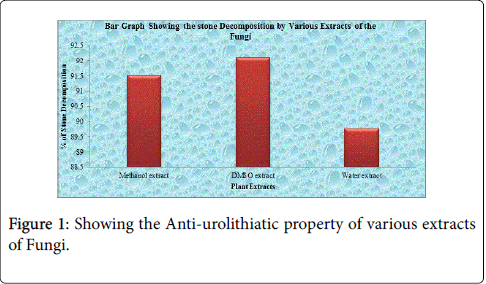
Figure 1: Showing the Anti-urolithiatic property of various extracts of Fungi.
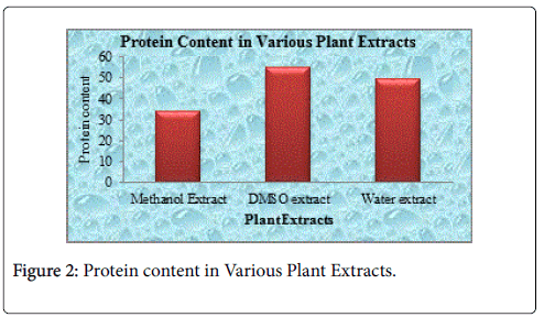
Figure 2: Protein content in Various Plant Extracts.
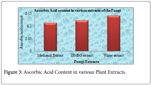
Figure 3: Ascorbic Acid Content in various Plant Extracts.
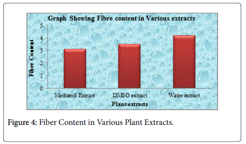
Figure 4: Fiber Content in Various Plant Extracts.
| S. No. | Plant Extract | Conc. of plant extract | Weight of calcium oxalate | Un-decomposed calcium oxalate | Decomposed calcium oxalate | % of oxalate decomposed |
|---|---|---|---|---|---|---|
| 1 | Methanol extract | 1000 mg | 1 | 0.0848 | 0.9152 | 91.52% |
| 2 | DMSO extract | 1000 mg | 1 | 0.079 | 0.921 | 92.10% |
| 3 | Water extract | 1000 mg | 1 | 0.1024 | 0.8976 | 89.76% |
Table 1: Showing the Anti-urolithiatic property of various extracts of Ganoderma lucidum.
| S. No | Plant extracts | Protein content (Conc. x 10-3 g g/l) |
|---|---|---|
| 1 | Methanol Extract | 34.3 |
| 2 | DMSO extract | 55.1 |
| 3 | Water extract | 49.5 |
Table 2: Showing protein content in various extracts of Ganoderma lucidum.
| S. No | Plant extracts | Ascorbic Acid content (Conc. x 10-3g g/l) |
|---|---|---|
| 1 | Methanol Extract | 0.111 |
| 2 | DMSO extract | 0.121 |
| 3 | Water extract | 0.14 |
Table 3: Showing the ascorbic acid content in various extracts of Ganoderma lucidum.
| S. No | Plant extracts | Fibre content (%) |
|---|---|---|
| 1 | Methanol Extract | 3.1 |
| 2 | DMSO extract | 3.5 |
| 3 | Water extract | 4.2 |
Table 4: Showing Fibre content in various extracts of Ganoderma lucidum.
Anti-arthritis activity of Ganoderma lucidium
The Anti-arthritis activity of various Ganoderma lucidium extracts Viz., (Methanol, DMSO and Water) have been analyzed, and it was found that (DMSO extract) (Figures 5-8) posses the highest antiarthritis activity followed by (water extract). The percentage inhibition by plant extracts was found to be concentration dependent, percentage inhibition increases with the increase in the concentration of the plant extracts. The IC50 value was determined from the straight line graph. The IC50 value of all the plant extracts was found lesser than the reference compound (acetyl salicylic acid). The IC50 value of various plant extracts follows the order (DMSO extract, water extract and inally Methanol Extract) was found to be (38, 50.5, 56) respectively and are presented in Tables 5-8.
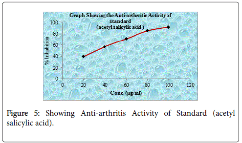
Figure 5: Showing Anti-arthritis Activity of Standard (acetyl salicylic acid).
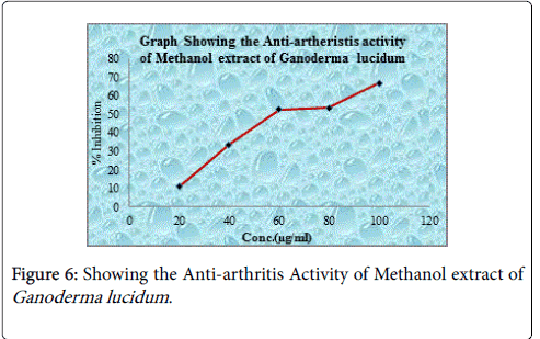
Figure 6: Showing the Anti-arthritis Activity of Methanol extract of Ganoderma lucidum .
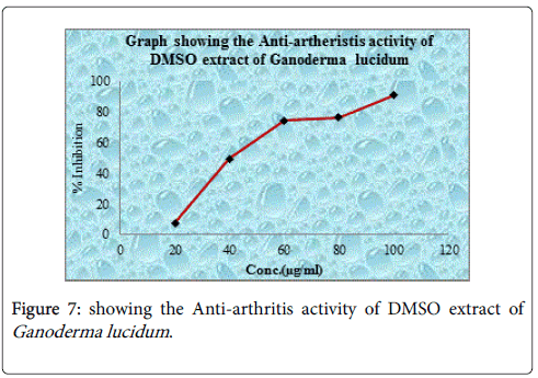
Figure 7: showing the Anti-arthritis activity of DMSO extract of Ganoderma lucidum .
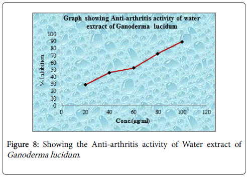
Figure 8: Showing the Anti-arthritis activity of Water extract of Ganoderma lucidum .
| S. No | Conc. µg/ml | Absorbance | % Red | IC50 Value |
|---|---|---|---|---|
| 1 | 20 | 0.545 | 39.61 | 32.5 |
| 2 | 40 | 0.456 | 57.12 | |
| 3 | 60 | 0.312 | 71.11 | |
| 4 | 80 | 0.198 | 85.43 | |
| 5 | 100 | 0.071 | 92.17 |
Table 5: Showing Anti-arthritis activity of acetyl salicylic acid Standard.
| S. No | Conc. µg/ml | Absorbance | % Red | IC50 Value |
|---|---|---|---|---|
| 1 | 20 | 0.855 | 11.81 | 56 |
| 2 | 40 | 0.651 | 34.62 | |
| 3 | 60 | 0.461 | 53.41 | |
| 4 | 80 | 0.451 | 54.35 | |
| 5 | 100 | 0.322 | 67.72 |
Table 6: Showing Anti-arthritis activity of Methanol extract of Ganoderma lucidum.
| S. No | Conc. g/ml | Absorbance | % Red | IC50 Value |
|---|---|---|---|---|
| 1 | 20 | 0.7825 | 38.14 | 38 |
| 2 | 40 | 0.442 | 48.27 | |
| 3 | 60 | 0.24 | 72.21 | |
| 4 | 80 | 0.212 | 71.43 | |
| 5 | 100 | 0.071 | 89.37 |
Table 7: Showing Anti-arthritis activity of DMSO extract of Ganoderma lucidum.
| S. No | Conc. g/ml | Absorbance | % Red | IC50 Value |
|---|---|---|---|---|
| 1 | 20 | 0.671 | 28.16 | 50.5 |
| 2 | 40 | 0.526 | 45.15 | |
| 3 | 60 | 0.445 | 51.5 | |
| 4 | 80 | 0.252 | 71.6 | |
| 5 | 100 | 0.105 | 88.27 |
Table 8: Showing Anti-arthritis activity of Water extract of Ganoderma lucidum.
The Ascorbic acid content of Methanol, DMSO and Water extracts were found to be (0.111, 0.121, 0.140 × 10⁻³g g/l). The protein content of Methanol, DMSO and Water extracts were found to be (34.3, 55.10, 49.5 x 10⁻³g/l). The fiber content of the concerned extracts was found to be 3.1, 3.5 and 4.2 respectively for Methanol, DMSO and Water Extract of the fungi.
The kingdom fungi is very interesting, because it furnishes all the types of metabolites which are very useful for the continuity of life, and for good health in addition helps in making our environment clean.
The fungi in relation showed a good response towards the presence of phytochemicals like, fibre, protein and ascorbic acid as per results is taken into consideration. It possesses alkaloids, terpenoids, saponins, flavonoids etc., differently in different plant extracts.
Protein denaturation or arthritis is a process in which proteins lose their tertiary structure and secondary structure by application of external stress or compound, such as strong acid or base, a concentrated inorganic salt, an organic solvent or heat. Most biological proteins lose their biological function when denatured. For example, enzymes lose their activity, because the substrates can no longer bind to the active site. Denaturation of protein is one of the cause of rheumatoid arthritis was documented. Production of auto antigen leads to denaturation of protein in certain arthritic disease. This antidenaturation effect was further supported by the change in viscosities. It has been reported that the viscosities of protein solutions increase on denaturation. From the result of the present study, it can be stated that all the extracts of Ganoderma lucidum are capable of controlling the production of auto-antigen and thereby it inhibit the denaturation of proteins of both fresh egg albumin and bovine albumin in dose dependent manner and its effect was compared with the standard drug diclofenac sodium. Water extract of Ganoderma lucidum have pronounced effect against arthritis, followed by DMSO and acetone extracts.
he work was performed by using in vitro anti-urolithiatic model for calculating percentage dissolution of kidney stone. This study has given primary evidence for Ganoderma as the fungi which possess lithiasis property. More specifically we can conclude from the results that Methanol and DMSO extracts both showing good anti urolithiasis activity. From the Table it is also clear that positive correlation exists between individual extracts and concentration used in the study. Out of various concentration used we can observe that activity increase as we increase the concentration and at one point further no increase in the activity observed. The fungi used in the above study also showing good activity when it was compared with the standard drug. The DMSO extract has been found to be more potent in terms of activity compare to Methanol and water. The above work recommends the fungi extract for the further studies by conducting in-vivo model.
The present set up of life, which is totally industrially based have so many ill effects against the human life. One among them is the stone problem in kidneys and in gall bladder, which is simply due to the utilization of chemical substances in the water bodies to kill microorganisms and other disease producing organisms. But these chemical substances by one way or another way find their entry into the human systems. Also among the humans the male generates are more prone to kidney stone related problems than the female generates, because of the biological differences in the two generates. In the presents efforts have been made to find some solution against the removal of kidney stone, whose ultimate cure is to operate a kidney for the removal of stone from it. The process of kidney operation is costly and is not out of danger. So from the present study many fungi extracts have been found cost effective against the removal of stone from the kidney, in which some measure have been made to make the breakdown of stone within the kidney and is then easily removed via urine.
Anti-arthritic effect of various extracts of Ganoderma lucidum was studied signi icantly by using in-vitro inhibition of protein denaturation model. The effect against the inhibition of protein denaturation was carried out by utilizing various extracts of Ganoderma lucidum . All the extracts had been found to be concentrations (dose levels) provided significant protection against denaturation of proteins. Most of the investigators have reported that denaturation of protein is one of the cause of rheumatoid arthritis. Production of auto-antigens in certain rheumatic diseases may be due to in vivo denaturation of proteins. Mechanism of denaturation probably involves alteration in electrostatic, hydrogen, hydrophobic and disulphide bonding. Obtained data stated that could be used as potent anti-arthritic agent.
As per the result obtained from the concerned fungi extracts, it was found that all the three fungi extracts bear a good nutritive value. In case where different solvents furnish different nutritive value is due to the interaction of these components with the concerned solvent differently, so different components have different type of concentrations in the respective solvents, which could be due to polarity index, hydrogen bonding and other mechanisms.
From the above results it could be concluded that all Ganoderma lucidum extracts showed a good response towards the fiber content. So the fungi in whole should be consumed regularly and as per the bodily demand as they boost up the bodily mechanisms by one way or another way. The secondary metabolites like fiber help in the maintenance of the primary metabolites. The ascorbic acid content, glucose and protein content in the three plant species is within the range of consumption, so are useful.
