Research Article - (2014) Volume 0, Issue 0
The methanol extracts of all the various parts of Morinda lucida were studied in vivo for anti-peroxidative, protective and ameliorative effects against carbon tetrachloride (CCl4) induced hepatocellular injury. Thiobarbituric acid reactive substances (TBARS) concentration taken as a measure of lipid peroxidation showed pre-treatment with the parts each (100 mg/kg) conferring anti-peroxidative effect while the leaf and bark extracts showed anti-peroxidative effect in the post-treatment test. Markers of hepatic damage viz: AST, ALT, total and conjugated bilirubin also showed significant protection and amelioration against CCl4-induced liver damage. Lipid profile studies like TG, TC, LDL and HDL showed no statistical difference among the pre-treatment groups and the ‘normal control’ except in the ‘Root+CCl4’ or ‘Leaf+CCl4’ groups which also showed no difference statistically from the ‘CCl4 only’ group. In the post-treatment (ameliorative) tests also, no statistical difference was found among the groups compared with the ‘normal control’ except the ‘CCl4 + Root’ and ‘CCl4 + Leaf’ groups which bore no statistical difference from the ‘CCl4 only’ group, as per LDL and HDL respectively. Findings here show the anti-peroxidative and protective properties of Morinda lucida comparable to Vitamins C and E, and ameliorative properties comparable to Silymarin against liver injury. These could possibly be reasons supporting its application in folkloric medicine.
Keywords: CCl4 ; Morinda lucida; Methanol extracts; Liver injury
Reactive oxygen species (ROS) are major sources of primary catalysts that initiate oxidation in vivo and in vitro which creates oxidative stress resulting in numerous diseases and disorders [1,2] such as cancer [3], cardiovascular disease [4], neural disorders [5], Alzheimer’s disease [6] mild congnitive impairment [7], Parkinsons disease [8], alcohol induced liver disease [9], ulcerative colitis [10], ageing [11] and atherosclerosis [12].
The extreme toxicity of oxygen is related to its high capability of generating free radicals which in turn destroys many major biological molecules. They can attack lipids and proteins and destroy membranes. Oxidized cellular thiols abstract hydrogen atoms from unsaturated fatty acids to initiate the peroxidation of membrane lipids [13]. A potent scavenger of these ROS could therefore serve as possible preventive intervention for free radical-mediated diseases [14].
Today, the search for natural compounds and other preparations of plant origin that can be used to prevent or mitigate maladies have been intensified. For instance, some medicinal and food plants such as Moringa oleifera [15], Khaya senegalensis [16], Syzygium aromaticum [17], Hibiscus esculentus [18], Canarium schweinfurthii [15] have been demonstrated not only to contain phytochemicals with strong antioxidant capacity, but possess diverse powerful medicinal properties.
Morinda lucida Benth, a Rubiaceae, is known commonly as ‘brimestone tree’ (English), ‘Arbre à soufre’ (French), and ‘Moindo’ (Polish). In Nigeria, it is called ‘oruwo’ or ‘erewo’ (Yoruba), ‘nfia’ or ‘Ezeogu’ (Igbo), Idonzakara (Hausa),‘ogele’ (Igala), among other many different indigenous tribal names, while in other African countries it is called ‘Uhon’ (Ivory Coast) and ‘hojologbo’ (Sierra Leone) [19-21]. It grows in many African countries and it is widely used as a medicine in West Africa. The plant is generally used as ingredients of fever teas, which are usually taken, for the traditional treatment of malaria [22]. The decoction of the stem bark has been reportedly used for the treatment of severe jaundice [23].
This study was thus designed to evaluate the antioxidant effect of the different parts of Morinda lucida and to determine the hepatoprotective/hepatocurative effects of the extract of the different parts of Morinda lucida on the integrity of the liver and associate disorders like hyperlipidemia and hyperbilirubinaemia.
Chemicals and reagents
All chemicals and reagents (of analytical grade) were obtained from BioVision Inc. (Milpitas, USA), Sigma Aldrich (Germany) or RANDOX Laboratories Randox® Ltd. (Ardmore, United Kingdom).
Experimental animals
Male albino rats of Wistar strain weighing between 180-280 g were used. They were obtained from Afkol farms, Jos, Plateau state and housed in Bingham University Animal facility. They were allowed to acclimatize for at least two weeks under the laboratory condition before the experiment was commenced. Except where and when applicable, animal were maintained on standard regular rodent pellet for rats procured from Vital Feeds depot, Gizzard plaza, Mararaba, Nasarawa State. All other protocols for animal management were observed in compliance with the general guidelines for animal experimentation.
Collection and authentication of plant sample
The different parts of Morinda lucida plant were collected from Idah Local Government Area of Kogi State. They were compressed in a plant press and sent to the Herbarum Unit of the Department of Biological Sciences, Ahmadu Bello University, Zaria for authentication. A specimen with voucher number: 1862 was deposited at the herbarium.
Plant extracts preparation and determination of percentage yield
Fresh plant parts, namely, whole fruit, leaf, bark, and root were collected and shade dried until brittle then pulverized with a mortar and pestle. Powders (25 g each) were then defatted with petroleum ether. Extraction was carried out with three changes of methanol for 5 hours each. The extracts were then pooled together and concentrated with rotary evaporator at 60°C for 3 hrs, then further allowed to dry over water bath at 45°C for 12 hrs. The pasty extracts were then transferred to desiccators containing activated silica gel for between 6-8 weeks until extracts became dry, depicted by constant weights recorded during the weekly weight measurements. Extracts yield were then quantified by method described by Mishra [24].
Animal groupings and treatments
Animals were broadly and randomly divided into various groups of 6 rats each. Groups I-VI served as the normal, CCl4 (negative control), fruit, leaf, bark or root only groups respectively. The test groups involved the extract pre-treatment (Hepatoprotective effect) and extract post-treatment (Ameliorative effect) groups. To investigate the former, animals received oral administration of 0.20 mg/kg Vitamin E (α-Tocopherol) [25,26] or 0.86 mg/kg Vitamin C (Ascorbic acid) [doses as low as 0.3 mg/kg [27], 30 mg/kg/day [28], and between 50- 200 mg/kg have been effectively reported to be used previously] or 100 mg/kg/day of extract (fruit, leaf, bark or root) on days 1 to 7 followed by 0.60 ml/kg CCl4 injection i.p., 30 min later (on day 7 only), in groups VI -XII respectively. In the latter, animals first received 0.60 ml/kg CCl4 injection i.p. on day 1, followed 30 min later by oral administration of 100 mg/kg of silymarin [29,30] or 100 mg/kg of fruit, leaf, bark or root extract on day 1, in groups XIII-XVII respectively. Afterwards, they received either silymarin or their respective extracts on days 2-7.
Collection of animal samples
Rats were sacrificed after administration of the methanol extracts of the fruit, leaf, bark and root of M. lucida for 7 days orally, and single dose of CCl4 injection (intra-peritoneally) on the first or last day for the pre or post treatment groups respectively. On the 7th day, the rats were allowed to fast overnight and then sacrificed under chloroform anesthesia by decapitation. Blood samples for determination of serobiochemical parameters were immediately collected.
Preparation of serum
Sera were prepared by allowing the collected blood to stand for 1 hour to clot. This was thereafter centrifuged at 1,000xg for 15 min to separate the serum which was harvested and stored in a freezer. The serobiochemical parameters studied were Asparate aminotransferase (AST), Alanine aminotransferase (ALT), total bilirubin, high density lipoprotein (HDL), low density lipoprotein (LDL), total cholesterol, and triglyceride concentration. At necropsy, all rats were examined to identify gross lesions, and the specimen of the liver were quickly removed and fixed in 10% formol-saline for histopathological studies.
Preparation of tissue homogenates
Liver tissues were removed immediately after sacrifice of rats, and washed in ice cold normal saline. They were then blotted, weighed and homogenized in 10 vols of ice cold PBS buffer (pH=7.4) followed by centrifugation of homogenate at 11,180xg for 30 min (at 4°C). The supernatant was stored frozen until used for the determination of TBARS [31-33], usually within 24 hrs.
Determination of lipid peroxidation
The assay is based on the reaction of thiobarbituric acid reacting substances (TBARS) with thiobarbituric acid (TBA) to form a pink coloured product. The colour intensity measured at 535 nm is directly proportional to TBARS concentration in the sample. Details of procedure were as previously described by Atawodi [17].
Determination of lipidemic parameters
Total serum triglyceride (TG), total cholesterol (TC), high density lipoprotein (HDL) and low density lipoprotein (LDL) concentrations were determined as per manufacturer’s instructions using assay kits (by Randox®).
Determination of liver function
Hepatotoxicity was induced in rats by method described by Gyamfi et al. [32] where single dose of 0.6 ml/kg of tetrachloromethane (CCl4) was injected i.p. AST and ALT activities were determined as per manufacturer’s instructions using assay kits (by Randox® Laboratories).
Estimation of bilirubin concentrations: Conjugated and total bilirubins were estimated as described by Garber [34] using respective assay kits (by Randox® Laboratories).
Analysis of Data: Data were expressed as mean ± SD. Significance of difference was evaluated by ANOVA and Duncan Multiple Range Test.
Protective influence of different parts of Morinda lucida on CCl4-induced oxidative stress and liver injury: Assay results showed no significant difference (p<0.05) in TBARS concentration between the normal control group, those pre-treated with α-Tocopherol or ascorbic acid or methanol extracts of the different parts of M. lucida before CCl4 injection, nor with the groups administered with the methanol extracts only. However, rats group injected with ‘CCl4 only’, was statistically higher than the normal control and all other groups (p<0.05) (Table 1). Similar to TBARS, markers of hepatic damage viz: AST, ALT, total and conjugated bilirubin showed no statistical difference among all the extract pre-treated groups and experimental controls except in the ‘CCl4 only’ group where values were significantly higher than all others. Histomicrographs of rats’ tissues attests to these (Figures 1 and 2).
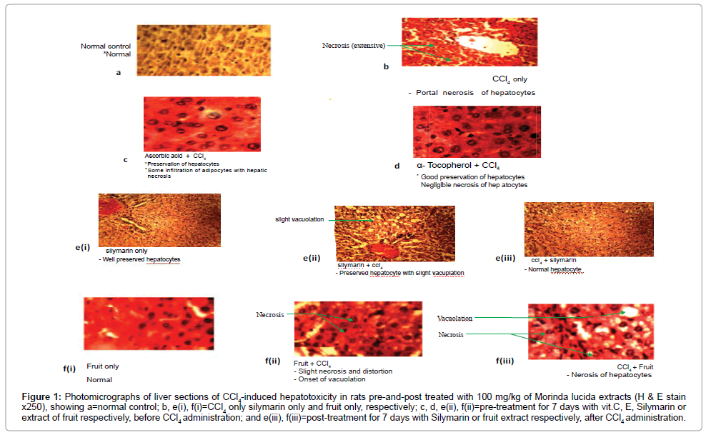
Figure 1: Photomicrographs of liver sections of CCl4-induced hepatotoxicity in rats pre-and-post treated with 100 mg/kg of Morinda lucida extracts (H & E stain x250), showing a=normal control; b, e(i), f(i)=CCl4 only silymarin only and fruit only, respectively; c, d, e(ii), f(ii)=pre-treatment for 7 days with vit.C, E, Silymarin or extract of fruit respectively, before CCl4 administration; and e(iii), f(iii)=post-treatment for 7 days with Silymarin or fruit extract respectively, after CCl4 administration.
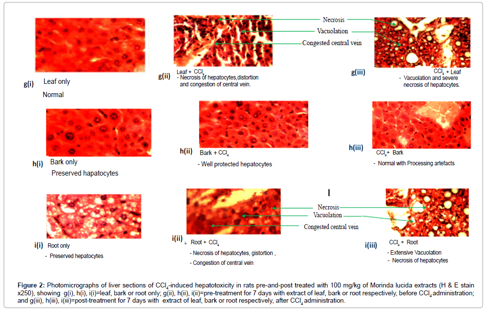
Figure 2: Photomicrographs of liver sections of CCl4-induced hepatotoxicity in rats pre-and-post treated with 100 mg/kg of Morinda lucida extracts (H & E stain x250), showing g(i), h(i), i(i)=leaf, bark or root only; g(ii), h(ii), i(ii)=pre-treatment for 7 days with extract of leaf, bark or root respectively, before CCl4 administration; and g(iii), h(iii), i(iii)=post-treatment for 7 days with extract of leaf, bark or root respectively, after CCl4 administration.
Values of lipidemic statuses like triglyceride (TG) concentration, total cholesterol (TC) and low density lipoprotein (LDL), showed ‘CCl4 only’ group being significantly higher (p<0.05) than the normal control group and those pre-treated with α-Tocopherol or ascorbic acid or methanol extracts of the different parts of M. lucida before CCl4 injection except in the ‘Root+CCl4’ and ‘Leaf+CCl4’ where there was no difference found compared to the ‘CCl4 only’ group (Table 2). HDL concentration showed significantly lower values (p>0.05) in the ‘CCl4 only’ group than the normal control and all other groups (Table 2, Figures 1 and 2).
Ameliorative influence of different parts of Morinda lucida on CCl4-induced oxidative stress and liver injury: No significant difference (p<0.05) in TBARS concentration among the normal control group and those post-treated with either silymarin or methanol extracts of the leaf or bark of M. lucida after CCl4 administration, but significantly lower than the fruit or root post-treated and the ‘CCl4 only’ groups (p>0.05) (Table 1). Markers of hepatic damage viz: AST, ALT, total and conjugated bilirubin showed no statistical difference between the normal, all the extract post-treated groups after CCl4 administration and experimental controls except in the ‘CCl4 only’ group where values were significantly higher (p<0.05) than all the others groups. These were also indicated in the histomicrographs (Figures 1 and 2).
Lipidemic statuses viz: triglyceride (TG) concentration, total cholesterol (TC) and low density lipoprotein (LDL), showed ‘CCl4 only’ group being significantly higher (p<0.05) than the normal control group, the silymarin group, and those post-treated with methanol extracts of the different parts of M. lucida after CCl4 administration except in the ‘CCl4+Root’ group, with respect to LDL concentration (Table 2). In HDL concentration, there was significantly lower values (p>0.05) in the ‘CCl4 only’ group than the normal control and all other groups except in the ‘CCl4+Leaf’ group (Table 2, Figures 1 and 2).
Treatments with plant extracts in hepatotoxicity studies for seven days have shown to be enough time to express hepatoprotective or ameliorative effect on induced liver damage [35-37]. In the hepatoprotective experiment, the C (water soluble) and E (lipid soluble) vitamins were used as the standard compounds due to their known roles in conferring protection against lipid peroxidation [38,39]. Since they are basically vitamins which are supplied to the body through diets, they offer protective effects against metabolic insults even before an ailment is fully established. Silymarin was employed as standard drug owing to its role as drug widely employed in the treatment of hepatic function impairment [40]. Silymarin, has been extensively studied and has shown to possess anticancer properties. It also has antioxidant properties, anti-metastatic activity and tumor suppressive effect on hepatocarcinogenesis [30,41].
From the acute toxicity study in rats using all the different parts of Morinda lucida, it was observed that the methanol extracts of parts of the plant were non-toxic, as indicated by the absence of any observed adverse effect even at the highest dose of 5000 mg/kg. According to Lorke [42], such compounds are considered non-toxic. Thus, the minimal 100 mg/kg/day dose of the methanol extracts of the fruit, leaf, bark and root of Morinda lucida was selected primarily, to ascertain potency, and also based on previous studies [43].
In this study also, the concentration of reactive oxygen species (ROS) measured as thiobarbituric acid (TBARS) concentrations, which is an index of lipid peroxidation [33] was increased in ‘CCl4 only’ rats group but significantly decreased in all the rats pre-treated with the different parts of M. lucida extracts. Also, among the rats treated with the different parts of extracts of M. lucida after administration of CCl4, there was a significant decrease in the MDA concentration among the groups that received leaf or bark extracts. While free radical chain reaction is widely accepted as a common mechanism of lipid peroxidation [39] which results in destruction of cell structures, lipids, proteins and nucleic acids [44], there was a clear evidence of the intervention of the extracts against the generation of free radicals.
Considering hepatoprotection/amelioration, there is replete of literature that serum enzymes particularly, alanine aminotransferase (ALT) and aspartate aminotransferase (AST) present in hepatic and biliary cells [45] are usually released from the hepatocytes and leak into circulation causing increase in their levels in the serum under hepatocellular injury or inflammation of biliary tract cells [45-47]. Hence, the elevated levels of ALT and AST are generally attributed to injury caused to the hepatocytes by carbon tetrachloride which affected the normal functions of the liver, since these enzymes are indisputably, markers of liver injury.
The determination of serum bilirubin is also an important marker for the diagnosis of several diseases, particularly high levels of bilirubin are strongly associated with hemolysis, blockage of the biliary tract, and liver disease [48]. Serum bilirubin (conjugated and total) levels were significantly elevated by CCl4 injection (Table 2). However, these were remarkably reduced by treatment with the methanolic extracts of various parts of M. lucida before and after CCl4 injection, suggesting the capabilities of the extracts to confer both hepatoprotection and amelioration, comparable to silymarin (Table 2, Figures 1 and 2). These findings have shown that the methanol extracts of M. lucida protected the rat livers against CCl4-induced damage and lowered the serum bilirubin in vivo.
With respect to lipid profile, evidence of significant decrease in triglyceride, total cholesterol and LDL concentrations among the hepatoprotective and ameliorative rat groups indicated a strong triglyceride and cholesterol lowering effect of the extracts except the leaf and root extracts in the hepatoprotective and ameliorative groups respectively (Table 2, Figures 1 and 2). Triglyceride, total cholesterol and LDL concentration among the ‘CCl4 only’ group was significantly very high, indicating the establishment of the hypercholesterolemic effect of the hepatotoxin. The observed increase in HDL among the rat groups among the hepatoprotective and ameliorative groups indicated the hyperlipidemia-lowering effect of the different methanol extracts of M. Lucida except in the ‘CCl4+leaf’ (ameliorative) group where values were similar to the ‘CCl4 only’ group as shown by the significantly low HDL concentrations due to the effect of CCl4 (Table 2). Like other plants constituents [49] the methanol extracts of parts of M. lucida (particularly the bark and leaf) reduced TG level. It could thus, be suggested that these extracts increased lipase activity which hydrolyzed TG. Among the lipids, increased blood level of TC, LDL and VLDL as well as lowered level of HDL have been identified as contributors in the development of hyperlipidemia [50] which is as a result of pathological conditions like liver disease and diabetes mellitus [51-53]. M. lucida may have acted as inhibitor for enzymes such as hydroxyl-methylglutaryl- CoA reductase, which is the key enzyme in de novo cholesterol biosynthesis [54,55]. This reduction could be beneficial in improving lipid metabolism and complications arising from same [56-62]. The present investigation has shown that extracts of parts of M. lucida improved liver synthetic activity.
There were reduced levels of thiobarbituric acid reactive substances (TBARS), ALT and AST, and concentrations of conjugated and total bilirubin, triglycerides, LDL and total cholesterol but higher HDL in the sera of animals that received CCl4 either before or after 7 days of administration of the various extracts particularly the bark and leaf. All these indicated the anti-peroxidative, hepatoprotective/ameliorative effects of these extracts on the integrity of the liver and associated disorders like hyperlipidemia and hyperbilirubinaemia. The findings in this work suggest that Morinda lucida, with particular reference to the leaves and bark could be utilized as important sources of natural antioxidants with health benefiting properties, and could be useful as additives in food or in nutraceuticals and functional foods and in biopharmaceutical industries.
