Research Article - (2018) Volume 6, Issue 1
Received Date: Nov 20, 2017 / Accepted Date: Dec 01, 2017 / Published Date: Dec 07, 2017
Salvinorin A, which is present in the natural plant Salvia divinorum, is a unique non-nitrogenous compound with hallucinogenic activity. When salvinorin A isolated from leaves of Salvia divinorum was irradiated with 300 nm UV light in ethyl acetate, it degraded from 100 μg/mL to 2.84 ± 0.05 μg/mL in 30 min. The calculated average rate constant k of this degradation was 0.12/min and the half-life was 5.7 min. When authentic salvinorin A was irradiated by UV light in an organic solution or an aqueous solution, it degraded over 90% within 40 min, whereas when it was irradiated by natural sunlight, it took 8 h to degrade 50% both in an organic and an aqueous solution. Three photodegradation products were tentatively identified by gas chromatography/mass spectrometry. Their structures were similar to that of salvinorin A, suggesting that they are also candidate non-nitrogenous hallucinogens.
Keywords: Salvia divinorum; Salvinorin A; Hallucinogens; Photodegradation
Salvia divinorum is a rare plant species that grew in obscurity until its accidental discovery in 1962 [1]. Growth occurs through branching of dangling nodes and then through rooting at the nodes, creating the bush-like appearance of the plant, which has been known to grow up to a maximum of three meters in height [2,3]. In early 1980, salvinorin A was discovered in this plant as a psychoactive component [4,5]. Salvinorin A is a unique chemical as it is the only non-nitrogenous hallucinogen and psychoactive diterpene [6] because hallucinogens occurring in natural plants, such as mushrooms, are generally nitrogen-containing compounds [7]. Later, several salvinorin A related compounds, such as salvinorin B, salvidivins A, and salvinicin A were identified in Salvia divinorum ; these are also unique to the plant [8,9]. These chemicals have been candidates for the psychoactive component of the plant because their structures are similar to that of salvinorin A. However, among these chemicals, only salvinorin A possesses significant hallucinogenic activity [5], suggesting that salvinorin A is the compound responsible for the hallucinogenic effect of Salvia divinorum [10].
Traditionally, salvia was used by the native Mazatec Indians from Oaxaca, Mexico, for divination, medico-religious ceremonies, and for its psychoactive effects [11]. In current times Salvia divinorum can be called salvia, magic mint, sally d, diviner’s sage, or Purple Sticky®, a particular brand [12]. Use of salvia is increasing as an alternative to marijuana [13,14]. Due to the dangers of smoking Salvia divinorum it has become illegal in several countries as well as in some states in the U.S. In order to establish regulations on the use of salvia to protect users, there is a pressing need to investigate various aspects of its nature, including its fate in the environment and its effects on humans after ingestion. Like many pharmaceutical compounds and illicit drugs, salvinorin A is unstable in the presence of light, but there are not many studies about salvinorin A and much is still unknown about it. Of particular import, there is no detailed information on its degradation pathway. In the present study, the behavior of salvinorin A under simulated and natural sunlight was investigated and tentative photo-degradation pathways of the products yielded was proposed.
Chemicals and materials
Salvinorin A (purity over 99%) was purchased from Sigma-Aldrich (Saint Louis, MO, USA). It was also bought online from Cayman Chemical Company (Ann Arbor, MI, USA). HPLC grade acetone, dichloromethane, ethyl acetate, and methanol were purchased from Fisher Scientific (Fair Lawn, NJ, USA). Salvinorin A was also isolated from dried Salvia divinorum leaves, which were purchased from SalviaSupply.com (Road Town, Virgin Island, UK).
Isolation of salvinorin A from Salvia divinorum
Dried Salvia divinorum leaves (20 g) were ground to a coarse power in a blender and then extracted twice with 1 L of acetone using a 2 L separatory funnel. After the acetone was removed by evaporation, the residual materials were transferred into a 200 mL separatory funnel containing 50 mL each of deionized water and ethyl acetate. The separatory funnel was shaken for 5 min and then the ethyl acetate layer was placed into a rotary evaporator to concentrate down to approximately 5 mL in volume. After the ethyl acetate solution was dried over minimum amount of anhydrous sodium sulfate. It was placed onto a 40 cm × 4.5 cm i.d. glass column packed with silica gel and eluted with an acetone/dichloromethane (5/95) solution. Over twenty fractions of 10 mL each were collected and analyzed for the presence of salvinorin A using gas chromatography/mass spectrometry (GC/MS). Identification of salvinorin A was confirmed comparing the mass spectral fragmentation and GC retention time of authentic salvinorin A purchased from the commercial sources. Fractions found to contain salvinorin A were condensed and recrystallized using methanol to provide a 95% pure salvinorin A.
Photo-irradiation of salvinorin A from Salvia divinorum with a photo-reactor
An ethyl acetate solution of salvinorin A (100 μg/mL) was placed into 8 quartz tubes (5 mL each), which were subsequently irradiated with UV light (λ=300 nm) in a Rayonet RPR-100 photochemical reactor (Branford, CT, USA) equipped with 163000 RPR UV bulbs. A tube was removed at each of the following time intervals, 1, 3, 5, 10, 15, 20, 25, and 30 min. Each reaction solution was analyzed for salvinorin A with the GC/FID.
Also, seven quartz tubes were filled with 4.2 mL each of aqueous solution (20 μg/mL) and irradiated with UV light. Tubes were removed after 5, 10, 20, 30, and 40 min of light exposure. An aqueous reaction solution from each tube (4 mL) was added to a Bond Elut® C-18 solid phase extraction (SPE) cartridge (Varian, Lake Forest, CA, USA) and eluted with 3 mL ethyl acetate. The resulting effluent was analyzed for salvinorin A using the GC/FID. The cartridges were conditioned with 5 mL each of deionized water, methanol, and ethyl acetate in series prior to use.
Salvinorin A is not directly soluble in water. Therefore, 4.0 mg of salvinorin A was originally dissolved in 10 mL methanol to get a 400 μg/mL solution. To create a 20 μg/mL solution in deionized water, 2.5 mL of salvinorin A solution in methanol was diluted to 50 mL with deionized water.
Recovery efficiency test on salvinorin A from solid phase extraction (SPE)
Three concentrations (80, 100, and 150 μg/mL) of 50/50-water/acetonitrile solution of standard salvinorin A were prepared and each solution was placed on a SPE cartridge of the type used above. After the water/acetonitrile was eluted, the cartridge was eluted with 5 mL ethyl acetate to recover salvinorin A. The ethyl acetate eluates were analyzed for salvinorin A by gas chromatography.
Photo-irradiation of salvinorin A with natural sunlight
An ethyl acetate solution (150 μg/g) of salvinorin A (1 mL) was placed in 24 quartz tubes. One test tube was wrapped with aluminum foil and used as a control. One tube was removed every 1 h and analyzed for degradation products. The experiment was carried out on October 8, 2014, on the roof of Meyer Hall at the University of California, Davis. The samples were exposed from 9 am to 5 pm. The weather was sunny and clear throughout the day.
Also, 1 mL each of a solution (water/acetonitrile=50/50) containing 100 μg/g salvinorin A was placed in 24 quartz test tubes. The control test tube was wrapped with foil. All tubes were exposed to natural sunlight and analyzed in triplicate. Test tubes were removed every hour. The experiment was carried out on February 24, 2015, on the roof of Meyer Hall at the University of California, Davis. The samples were exposed from 9:15 am to 5:15 pm. The weather was sunny and clear. After irradiation, the samples were prepared in ethyl acetate solution using the SPE described above for GC/MS analysis.
Instrumental
The irradiated samples were analyzed for salvinorin A by an Agilent 6890 series gas chromatograph coupled with a flame ionization detector (GC/FID). The GC was equipped with a 15.0 m × 250 μm × 0.1 μm DB-1 fused silica capillary column (Aligent, Folsom, CA, USA).
Helium carrier gas flow rate was 1.0 mL/min. The GC oven temperature was held at 150°C for 1 min and then programmed to rise to 250°C at 30°C/min. The injector and detector temperatures were 300°C and 280°C, respectively.
All standards and irradiated samples were also analyzed by an Agilent Technologies 6890 N Gas Chromatograph interfaced to an Agilent Technologies 5973 Network Mass Selective Detector (GC/ MSD). The column used in the GC system was a 30 m × 250 μm × 0.25 μm DB-1 fused silica capillary column (Aligent, Folsom, CA, USA). Helium carrier gas flow rate was 1.0 mL/min. The inlet and the GC/MSD interface were set to 250°C and 280°C, respectively. The initial oven temperature was 80°C and was held for 2 min, then programmed to rise to 280°C at 20°C/min. The final temperature was held for 8 min. The electron energy of the MS was 70 eV. The quadrupole mass spectrometer was set to scan from 50 to 550 m/z. The temperatures of the ionization source and the quadrupole were 230°C and 150°C, respectively.
Tentative identification of photo-degradation products was performed using our knowledge for elucidation of structures based on MS fragmentation. Also, the NIST02 mass spectral library (National Institute of Standards and Technology, Gaithersburg, MD, USA) and salvinorin A related compounds reported previously [15] were referred to for further confirmation of our identifications.
As mentioned above, preparation of an aqueous salvinorin A is not easy because of its insolubility in water. However, salvinorin A was readily dissolved in a solution of water/acetonitrile (50/50). The preliminary experiments on photo-irradiation of salvinorin A in aqueous and water/acetonitrile solutions showed no significant difference. Therefore, a water/acetonitrile solution was used for further photo-degradation experiments.
The limit of detection (LOD) and the limit of quantitation for salvinorin A in an ethyl acetate solution determined according to a previously reported method [16] were 7.02 μg/mL and 23.4 μg/mL, respectively. These values are relatively high, but represent a very conservative estimate, since the instrument could actually produce a measurable peak at a concentration of 0.50 μg/mL in the present study.
The recovery efficiencies of SPE used were 79.82 ± 0.80% for 80 μg/mL, 99.78 ± 1.94% for 100 μg/mL, and 94.35 ± 3.73% for 150 μg/mL from a water/acetonitrile solution.
Figure 1 shows the photo-degradation of salvinorin A isolated from the leaves of Salvia divinorum in ethyl acetate irradiated with 300 nm UV light for 30 min. Values are mean ± SD (n=3). SD ranged from ± 5.32 (3 min irradiation) to ± 0.03 (25 min irradiation) but the values less than ± 2.30 do not appear in Figure 1. The degradation occurred rather quickly. Salvinorin A (100 μg/mL) was degraded down to 2.84 ± 0.05 μg/mL after 30 min. The calculated average rate constant k of this degradation was 0.12/min and the half-life was 5.7 min. The degradation rate constant and half-life were calculated using the following first-order reaction equations [17]:
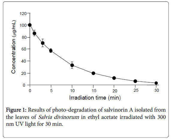
Figure 1: Results of photo-degradation of salvinorin A isolated from the leaves of Salvia divinorum in ethyl acetate irradiated with 300 nm UV light for 30 min.
In[Salvinorin A]t/[Salvinorin A]0=-kt and t1/2=In2/k
Where [Salvinorin A]t represents the concentration of salvinorin A at time t, [Salvinorin A]0 represents the concentration of salvinorin A at time 0, k represents the rate constant, and t1/2 is the half-life. The rate constants were determined by plotting the graphs of In[Salvinorin A]t/[Salvinorin A]0 against time.
Figure 2 shows the photo-degradation of salvinorin A isolated from leaves of Salvia divinorum in water irradiated with 300 nm UV light for 40 min. Salvinorin A (20 μg/mL) was degraded down to 3.15 ± 0.01 μg/mL after 40 min. The calculated average rate constant of this degradation was 0.044/min and the half-life time was 16 min. When salvinorin A isolated from Salvia divinorum leaves was irradiated with 300 nm UV light in an ethyl acetate or water solution, it degraded almost three times slower in water than in ethyl acetate.
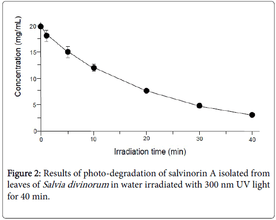
Figure 2: Results of photo-degradation of salvinorin A isolated from leaves of Salvia divinorum in water irradiated with 300 nm UV light for 40 min.
Figure 3 shows the photo-degradation of authentic salvinorin A in ethyl acetate and water/acetonitrile (50/50) solutions irradiated with 300 nm UV light for 40 min. Values are mean ± SD (n=3). In the case of the ethyl acetate solution, SD ranged from ± 4.95 (10 min irradiation) to ± 0.05 (20 min irradiation); values less than ± 2.60 do not appear in Figure 3. Authentic salvinorin A degraded from 100 μg/mL to 8.33 μg/mL after 30 min UV irradiation. The calculated average rate constant of this degradation was 0.8/min and the half-life time was 8.7 min.
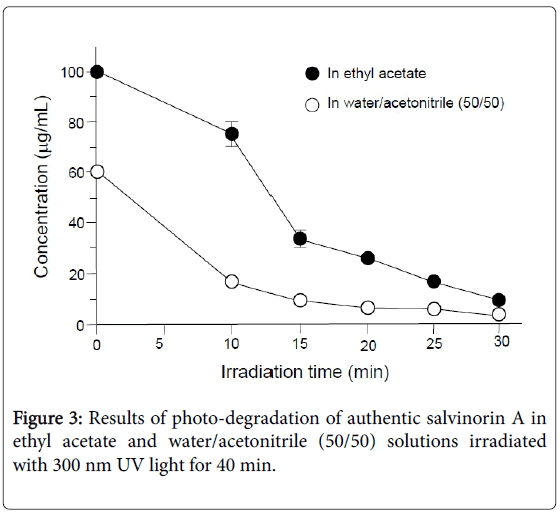
Figure 3: Results of photo-degradation of authentic salvinorin A in ethyl acetate and water/acetonitrile (50/50) solutions irradiated with 300 nm UV light for 40 min.
In the case of the water/acetonitrile (50/50) solution, SD ranged from ± 1.09 (10 min irradiation) to ± 0.01 (25 min irradiation); values less than ± 2.60 do not appear in Figure 3. Authentic salvinorin A degraded from 60 μg/mL to 2.86 μg/L after 30 min UV irradiation. The calculated average rate constant of this degradation was 0.09/min and the half-life time was 7.7 min.
Figure 4 shows the photo-degradation of authentic salvinorin A in ethyl acetate and water/acetonitrile (50/50) solutions irradiated with natural sunlight for 30 min. Values are mean ± SD (n=3). In the case of an ethyl acetate solution, SD ranged from ± 8.16 (5 h irradiation) to ± 2.51 (2 h irradiation); values less than ± 2.91 do not appear in Figure 4. Authentic salvinorin A degraded from 150 μg/mL to 71.89 μg/mL after 40 min sunlight irradiation. The calculated average rate constant of this degradation was 0.08/h and the half-life time was 8.84 h.
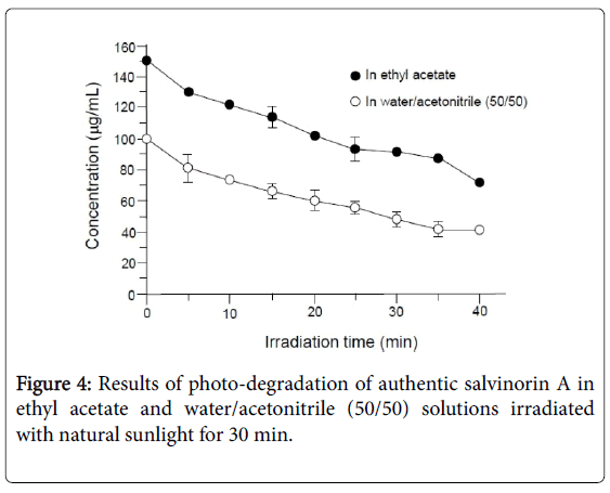
Figure 4: Results of photo-degradation of salvinorin A isolated from leaves of Salvia divinorum in water irradiated with 300 nm UV light for 40 min.
In the case of the water/acetonitrile (50/50) solution, SD ranged from ± 9.13 (5 min irradiation) to ± 2.54 (40 min irradiation); values less than ± 2.91 do not appear in Figure 4. Authentic salvinorin A degraded from 100 μg/mL to 40.03 μg/mL after 40 min sunlight irradiation. The calculated average rate constant of this degradation was 0.10/h and the half-life time was 6.82 h.
All figures exhibited similar patterns of first-order degradation curves. When salvinorin A from Salvia divinorum leaves was irradiated with 300 nm UV light in ethyl acetate or water, degradation occurred much faster in ethyl acetate than in water. This may be due to its lessened solubility in water because it degraded in water/acetonitrile solution almost as fast as in ethyl acetate. Also, Salvia divinorum leaves contain some water soluble chemicals which inhibit the photodegradation of salvinorin A. The results indicate that salvinorin A degrades readily by UV light, suggesting that it is important to prevent exposure to UV light for determination of the levels of salvinorin A in natural plants after collection.
The degradation rates calculated are at a rather accelerated rate based on high UV exposure. A study of the metabolism of salvinorin A in rat plasma found a degradation rate of 0.11/h and a half-life of 6.30 h at 25°C [18]. This study also found that increasing the temperature increased the rate of degradation with less than two percent of the original salvinorin A present in the sample after incubation at 37°C, or body temperature, for 24 h. However, the degradation rate of salvinorin A by a living organism is comparable to that of UV irradiation. The quick breakdown of salvinorin A in living organisms may account for the short duration of its hallucinogenic effects.
Figure 5 shows a typical gas chromatogram of a sunlight irradiated (20 min) sample in ethyl acetate. Refer to Table 1 for details of the degradation products tentatively identified. Gas chromatography has been used widely for analysis of plant extracts [19].
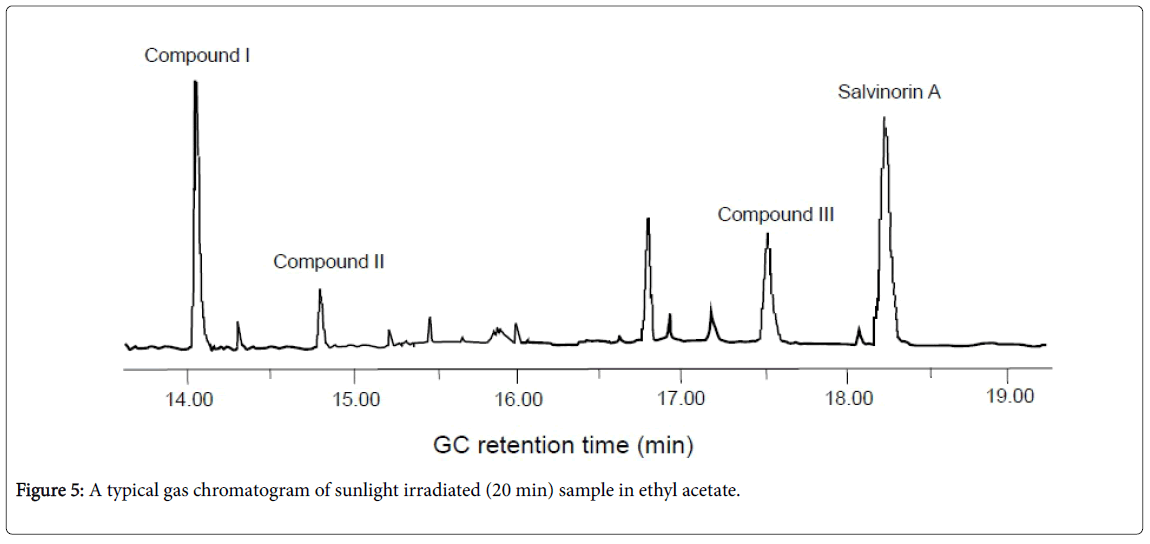
Figure 5: A typical gas chromatogram of sunlight irradiated (20 min) sample in ethyl acetate.
| Compound | Systematic name | GC retention time (min) | MS fragment, m/z (%) |
|---|---|---|---|
| Salvinorin A | methyl (2S,4aR,6aR,7R,9S,10aS,10bR)-9-(acetyloxy)-2-(furan-3-yl)-6a,10b-dimethyl-4,10-dioxo-dodecahydro-1H-naphtho[2,1-c]pyran-7-carboxylate | 18.26 | 432 (M+, 11.0), 404 (8.5), 359 (8.0), 318 (6.4), 273 (23.8), 220 (12.1), 166 (15.6), 121 (19.7), 94 (100), 43 (41.9) |
| Compound I | methyl (4S,4aR,5S,6R,8aR)-5-[2-(3-furyl)ethenyl]-4-hydroxy-6(hydroxymethyl)-5,8a-dimethyl-3,4,4a,5,6,7,8,8a-octahydro-1-naphthalenecarboxylate | 14.04 | 360 (M+, 33.1), 342 (6.4), 328 (8.3), 301 (85.7), 283 (18.6), 245 (37.6), 121 (100), 107 (56.6), 95 (80.8), 81 (58.0), 55 (31.6) |
| Compound II | methyl (6aR,7R,10bR)-2-(furan-3-yl)-6a,10bdimethyl-4,10 -dioxo-1,4,4a,5,6,6a,7,10,10a,10b-decahydro-2Hbenzo[f] isochromene-7-carboxylate | 14.80 | 372 (M+, 64.4), 340 (9.4), 312 (100), 297 (39.9), 253 (16.1), 173 (39.7), 159 (28.6), 145 (64.2), 131 (35.2), 105 (28.2), 91 (47.6), 55 (19.2) |
| Compound III | methyl (2R, E)-2-((4aR,6R)-5-formyl-3-(furan3-yl)-4a,6-dimethyl -1-oxooctahydro-1Hisochromen-6-yl)-4-(prop-1-en-2-yloxy) but-3enoate | 17.20 | 432 (M+, 15.1), 404 (25.8), 318 (12.6), 273 (79.6), 2.6 (33.4), 146 (28.6), 121 (47.5), 107 (22.5), 94 (100), 81 (13.2), 55 (26) |
Table 1: Photo-degradation products and salvinorin A identified in an ethyl acetate solution of authentic salvinorin A irradiated with 300 nm UV light.
Figure 6 presents proposed formation pathways of these compounds. Identification of salvinorin A in this sunlight irradiated solution was confirmed using an authentic chemical purchased from commercial sources. Compound II was proposed to form from salvinorin A via a Norrish type I photo-reaction cleaving a bond between carbon numbers 1 and 2. Intermediate A was formed via hemolytic cleavage between carbon number 2 and acetate oxygen and subsequently Compound II was formed upon losing a hydrogen radical. Compound I may form through intermediate B from Compound II as shown in Figure 6. However, formation of this compound may come from other products. A previous study reported the presence of a similar compound, which has a saturated bond between carbon numbers 11 and 12 [15]. They proposed the structure of this compound using the 1H NMR spectrum.
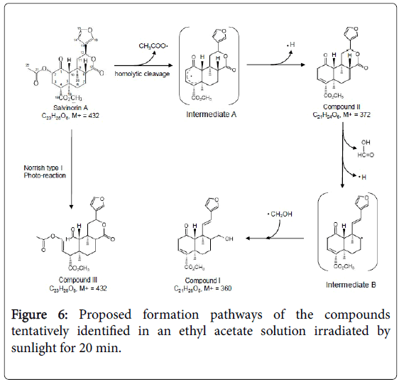
Figure 6: Proposed formation pathways of the compounds tentatively identified in an ethyl acetate solution irradiated by sunlight for 20 min.
Naturally occurring hallucinogens containing no nitrogen atom, such as salvinorin A, are unique. Photo-degradation of salvinorin A occurs in relatively short time, suggesting that it is necessary to store in appropriate light-free conditions. In addition, salvinorin A produces various photo-degradation compounds, which may possess hallucinogenic effects. In the present study, salvinorin A isolated from a natural plant, Salvia divinorum , behaved the same as the authentic chemical. Detailed study of the hallucinogenic activity of these photodegradation products is in order.
