Research Article - (2018) Volume 6, Issue 2
Received Date: Jan 05, 2018 / Accepted Date: Jan 17, 2018 / Published Date: Jan 25, 2018
Leaves of Plantago lanceolata were traditionally used to treat wounds, burns, inflammations, fevers, diabetes and cancer. The present study was carried out on the phytochemical investigation and antimicrobial activities of the leaves extract of Plantago lanceolata since the plant was used for wound healing in Ethiopia. The powdered leaves of Plantago lanceolata herb was sequentially extracted with organic solvents: petroleum ether, chloroform/methanol (1:1) and methanol respectively. The crude extracts was subjected to phytochemical screening and revealed the presence of steroids, alkaloids, flavonoids, saponins, glycosides, phenols, tannins and terpenoids compounds that might be responsible for the claimed activities by local people. The petroleum ether extract was purified over silica gel preparative thin layer chromatography and yielded an isolated compound PL-5. The structure of this compound was elucidated using different spectroscopic techniques such as FT-IR, 1H-NMR, 13C-NMR and DEPT-135 spectral data and by comparing the data with literature reports. The crude extracts, isolated pure compound and n-hexane extracted oil were tested against four bacterial species (Gram negative bacteria: Escherichia coli and Salmonela thyphei; Gram positive bacteria: Staphylococcus aureus, Streptococcus agalactiae) and two fungal species (Aspergillus niger and Fusarium solani) using paper disc diffusion method. All crude extracts, isolated pure compounds and extracted oil were active against all the tested bacterial. Additionally, petroleum ether and chloroform/methanol (1:1) crude extracts and n-hexane extracted oil were active against the two fungal species and hence the present work supported the medicinal use of Plantago lanceolata.
Keywords: Antimicrobial activities; Phytochemical; Screening; Plantago lanceolata
Medicinal plants are now more focused than ever because they have the capability of producing many benefits to society indeed to mankind, especially in the line of medicine and pharmaceutical. Plant parts such as leaves, roots and bark are used for the therapeutic purposes and as well serve as precursors for the synthesis of useful drugs due to their ethnomedical importance in nature. The medicinal potentials of these plants could be traceable to the bioactive phytochemical constituents that are responsible for the physiological action on the human body [1]. Substances derived from plants have recently being of great interest due to their versatility. These substances in the plant which enhance their usefulness globally are classified as phytochemicals [2].
Majority of the people living in the developing world are struggling to increase the standard of living and to improve the health care delivery in the face of increasing poverty and growing population. According to WHO survey, 80% of populations living in the developing countries rely exclusively on traditional medicine for their primary health care needs of which most involve the use of plant extracts [3]. Plantago lanceolata belonging to the Plantaginaceae family has also taken place in many medicinal uses as a wound healing remedy. This herbaceous plant was traditionally used in North Africa to treat wounds, burns, abscesses, inflammations, hemorrhoids and fevers [4]. Previous reports have indicated that the plant was also effective against diarrhea, dysentery, an anesthetic, antiviral, antiinflammatory, astringent, anti-helmintic, analgesic, analeptic, antihistaminic, anti-rheumatic, antitumor, anti-ulcer, diuretic, expectorant and hypotensive in traditional medicine [5,6]. Among many popular medicinal plants, Plantago lanceolata has accessed a scientific value, as it has been taken place in many traditional medicine uses as a wound healing remedy for centuries [7]. In this study, better extraction methods of Plantago lanceolata leaves were investigated. For effective extraction and better yield of extract of Plantago lanceolata oil, organic solvent extraction methods was used and certain parameters such as extraction time and temperature was manipulated.
Sample collection
The leaves of Plantago lanceolata L., herb was collected from Haramaya University main campus, Ethiopia, in December 2016 (Figure 1).
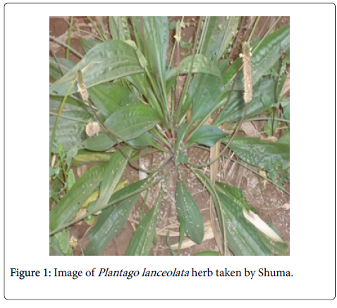
Figure 1: Image of Plantago lanceolata herb taken by Shuma.
The bacterial and fungal test microorganisms used in this study were two Gram positive bacterium: Staphylococcus aureus and Streptococcus agalactiae and two Gram negative bacterium: Escherichia coli and Salmonela thyphei and the fungi: Aspergillus niger and Fusarium solani were obtained from Plant Pathology Laboratory of the School of Plant Science, Haramaya University.
Sample preparation and crude extraction
The collected leaf was washed repeatedly and dried in an open air protected from direct exposure to sun light. Air dried leaves of Plantago lanceolata L., was grinded by analytical mill and packed in polyethylene bags. A 210 g of the powdered of P. lanceolata L., leaves was then extracted with petroleum ether for 8 h in soxhlet apparatus (60 g of each) and soaked with 300 mL of petroleum ether and heated with heating mantle at 45°C for 8 h and filtered with filter paper (Figure 2). The filtrate was collected and concentrated at 40°C under reduced pressure using a Rotary evaporator. After air drying at room temperature, the defatted marc was subjected soxhlet extractor with MeOH: CHCl3 (1:1) for 8 hrs. Then filtered using what man No.1 filter paper and concentrated using Rotary evaporator at 40°C under reduced pressure. The marc collected after CHCl3/MeOH extraction was dried at room temperature for further extraction with MeOH by the same procedure. This procedure was repeated twice until sufficient crude extract was collected and the crude extract was kept at 4°C until analysis.
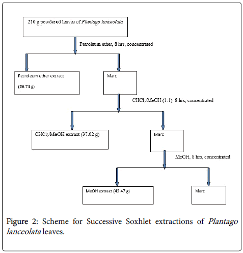
Figure 2: Scheme for Successive Soxhlet extractions of Plantago lanceolata leaves..
Yield of extract %=Weight of crude extract/Weight of samples × 100.
Phytochemicals screening on leaves of Plantago lanceolata
There was phytochemical screening of methanol crude extract report on the leaves of the Plantago lanceolata [8]. In this study, the preliminary phytochemical screening was carried out on the plant extract (petroleum ether crude extract, CHCl3/MeOH (1:1) crude extract and MeOH crude extract), following the standard procedures were described [9,10].
1. Detection of saponins:
Froth test: All extracts (0.25 g each) were diluted with distilled water to 10 mL and shaken in a graduated cylinder for 15 min vigorously. The foam formation indicated the presence of saponins.
2. Detection of quinones: 0.2 g of each of the Pet.ether, CHCl3/ MeOH (1:1) and MeOH crude extracts were treated separately with alcoholic potassium hydroxide solution. Then, the formation of blue color from red indicated the presence of quinones.
3. Detection of carbohydrates: Pet.ether, CHCl3/MeOH (1:1) and MeOH crude extracts were dissolved individually in 5 ml distilled water and filtered. The filtrates were used to test for the presence of carbohydrates.
Molisch’s solution test: Molish’s solution was added to the crude plant extracts dissolved in distilled water then 2 mL of H2SO4 concentrated was added and poured carefully along the side of the test tube, a violet ring appeared at the interphase of the test tube indicated the presence of carbohydrate.
4. Detection of flavonoids:
Lead acetate test: Pet.ether, CHCl3/MeOH (1:1) and MeOH crude extracts (0.2 g) were treated with few drops of lead acetate solution. Formation of yellow color precipitate indicates the presence of flavonoids.
5. Detection of alkaloids: 0.2 g of Pet.ether, CHCl3/MeOH (1:1) and MeOH crude extracts were dissolved separately in 5 mL diluted Hydrochloric acid and filtered.
Wagner’s test: 2 mL Filtrates were treated with Wagner’s reagent (Iodine in Potassium Iodide). Formation of brown/reddish precipitate indicates the presence of alkaloids.
6. Detection of tannins: 0.2 g of Pet.ether, CHCl3/MeOH (1:1) and MeOH crude extracts was dissolved in 10 mL distilled water and filtered. 3-4 drops of 1% aqueous Iron chloride (FeCl3) solution was added to the filtrate. The appearance of intense green, purple, blue or black color indicated the presence of tannins in the test samples.
7. Detection of phenols: Ferric Chloride Test: Pet.ether, CHCl3/ MeOH (1:1) and MeOH crude extracts (0.25 g) were treated with 3-4 drops of ferric chloride solution. Formation of bluish black color indicated the presence of phenols.
8. Detection of terpenoids: 0.2 g of the organic extract was dissolved in 2 mL of CHCl3 and evaporated to dryness. 2 mL of conc. H2SO4 was then added and heated for about 2 min. Development of a grayish color was indicated the presence of terpenoids.
9. Detection of steroids:
Salkowski’s test: The formation of red color in the lower chloroform layer when 0.2 g of organic extracts (Pet.ether, CHCl3/MeOH (1:1) and MeOH crude extracts) dissolved in 2 mL of chloroform and 2 mL concentrated sulphuric acid added to it, indicates the presence of steroids.
10. Detection of glycosides:
Keller-Kiliani Test: 0.2 g extracts of pet. Ether, CHCl3/MeOH (1:1) and MeOH was added to glacial acetic acid, one drop of 5% FeCl3 and conc.H2SO4. Reddish brown color was appeared at junction of the two liquid layers and upper layer appears bluish green indicated the presence of glycosides.
Structural elucidation
Characterizations of the isolated compounds were governed by spectroscopic techniques through the overdue conditions. NMR spectra were recorded on Brucker Avance DMX 400 FT-NMR spectrometer operating at 400 MHz for 1H and 100 MHz for 13C at room temperature by using CDCl3. Tetra methylsilane (TMS) was used to refer as standard. IR spectra were recorded between 400-4000 cm-1 in KBr pellets.
Isolation of compounds
Preparative thin layer chromatography and solvent system: Preparative Thin-layer chromatography (PTLC), glass plates admeasuring 20 × 20 cm were coated with silica gel G (1-7 mm). They were dried at room temperature for 5-6 hours and activated in oven at 110°C for almost the same period. After dried the band subjected to isolate through a chromatographic chamber using the overdue solvent system. Approximately 5-10 mg of sample (fractions) was dissolved in 1-2 ml Pet. Ether and applied over the activated silica layer as a narrow streak across the plate. The solvent from the streak was allowed to evaporate. The plate was then developed in toluene:ethylacetate (40: 10) binary mobile phase in a rectangular tank. After the development, the plate was removed from the tank and the solvents were allowed to evaporate at room temperature. The compound from the scraped silica was recovered by adding appropriate volume of Pet. Ether followed by shaking and filtration. The solute was thus extracted four times and the extracts were combined.
A mixture of toluene and ethyl acetate in the ratio of (4:1) was used as solvent systems to isolate six bands with Rf values of 0.17, 0.50, 0.58, 0.70, 0.78 and 1.00 and the purity of (Rf=0.78, 1.00) was checked on analytical TLC.
Coding system for isolated pure compound
The coding system of the isolated pure compound was based on the first two letters from the scientific name of the plant; P stands for the genus name Plantago , L stands for the place of specimen lanceolata and followed by the number that indicating the location of the compounds starting from the highest Rf value to the lowest. TLC examination of the crude extracts revealed the presence of at least six spots visualized under UV lamp at 254 nm and 356 nm. PL-1, PL-2 and PL-5 indicate the first, second and fifth compounds respectively.
Antimicrobial Assay
Preparation of inoculums: The test bacterial strains were transferred from the stock cultures and streaked on Mueller Hinton plates and incubated for 24 hrs at 37°C. Well separated bacterial colonies were then used as inoculums. Bacteria was transferred using bacteriological loop to autoclaved MHA that was cooled to about 45°C in water bath and mixed by gently swirling the flasks. The medium was then poured to sterile Petridishes, allowed to solidify and used for the Biotest [11].
For test fungi, mycelial plugs from stock cultures were transferred to PDA plates and incubated for 3 days. Then spores of A. niger was harvested by washing the surface of the colony using 10 mL sterile distilled water and was transferred to 250 mL autoclaved PDA which was cooled to about 45°C in water bath. Likewise, mycelium of F. solani was washed with 10 mL sterile distilled water, macerated in blander and the mycelia suspension was transferred to 250 mL autoclaved PDA cooled to about 45°C in water bath. The medium containing spore or mycelia suspension was poured to sterile a plates allowed to solidify and used for disk diffusion bioassay [11].
Testing for antifungal and antibacterial activity: A filter paper disc of about 6 mm in diameter was placed in beaker sterilized in an oven at 180°C for 1 hrs. 10 and 20 μL of the samples were pipetted to the discs in three replications. The paper discs impregnated with the extract solutions were then transferred using sterile forceps to PDA seeded with spore or mycelia suspension of test fungi as described under inoculums preparation above. The Petri dishes were incubated at 24°C for 3 days. The antifungal activity was evaluated by measuring of the inhibition zone against the tested organisms. The entire test was performed in triplicate. Similar procedures to that of antifungal test were followed. Sterilized paper discs were transferred to MHA plates seeded with at 37°C bacteria for 24 hrs. All the tests were performed in triplicate. The leaves crude extracts, fractions, n-hexane extracted oil and pure compounds were taken to test the sensitivity towards four bacteria.
Determination of extraction yield
The extraction yield is a measure of the solvent efficiency to extract specific components from the original material. The percentage yield of crude extract in respective solvent crude extract was shown in Table 1. It could be calculated according to the formula as follows [12].
| Crude extract | Yield in (%) |
|---|---|
| Petroleum ether | 7.97 |
| CHCl3/MeOH | 19.16 |
| MeOH | 27.18 |
Table 1: Yield of Petroleum ether, CHCl3/MeOH and MeOH crude extracts by Soxhlet extractor.
Percentage yield=Weight of crude extract/Weight of samples × 100
Phytochemical constituent
In the present study, the qualitative analyses of P. lanceolata leaf extracts were carried out for dried leaf samples. The preliminary phytochemical screenings on leaves (petroleum ether, CHCl3/MeOH and MeOH) extracts indicated the presence of saponins, tannins, alkaloids, flavonoids, terpenoids, and phenolic compounds were confirmed in leaves of extracts of P. lanceolata . The phytochemical screening test results of Pet.ether, CHCl3/MeOH and MeOH crude extracts of P. lanceolata leaves is presented in the Table 2 below.
| No | Constituents | Pet. Ether | CHCl3/MeOH | MeOH |
|---|---|---|---|---|
| 1 | Steroids | + | + | + |
| 2 | Alkaloids | + | + | - |
| 3 | Carbohydrates | - | - | - |
| 4 | Flavonoids | - | + | + |
| 5 | Saponins | - | + | ++ |
| 6 | Glycosides | - | + | + |
| 7 | Phenols | - | - | + |
| 8 | Tannins | - | + | ++ |
| 9 | Terpenoids | + | + | - |
| 10 | Quinones | - | - | - |
Table 2: Phytochemical screening results of P. lanceolata leaves.
The phytochemical screening test of Pet.ether crude extract showed that the presence of steroids, alkaloids, terpenoids and the absence of glycosides, carbohydrates, flavonoids, saponins, phenols, tannins and quinones. The CHCl3/MeOH crude extract has steroids, alkaloids, flavonoids, saponins, glycosides, tannins, and terpenoids and has no carbohydrates, phenols and quinones. The methanol extract has constituted steroids, flavonoids, saponins, glycosides, phenols, and tannins and has no constituted alkaloids, carbohydrates, terpenoids and quinones (Table 2).
Structural elucidation of the compound PL-5
Compound PL-5 was isolated as a purple reddish amorphous solid with an Rf value of 0.78 which was obtained from petroleum ether extract isolated by PTLC in toluene/ethyl acetate (40:10). Structural elucidation of the compounds was based on the spectroscopic data obtained from FT-IR, NMR (1H-NMR, 13C-NMR and DEPT-135).
The IR spectrum of compound PL-5 revealed a medium absorption band at 3433 cm-1 due to the presence of a stretching vibration of -OH group. The strong absorption band at 2922 and 2853 cm-1 is due to aliphatic CH3 and CH2 stretching vibration frequencies respectively. The absorption band at 1754 cm-1 indicates the presence of carboxyl groups whereas the absorption band at 1464 cm-1 and 693 cm-1 indicates the -CH2 bending for alkanes and -CH bending for unsubstituted alkene.
The 1H-NMR spectrum of PL-5 showed a downfield shifted at 5.314 ppm, due to the presence of olefinic proton (H-20). The spectrum showed the presence of methyl proton at δH 0.81-1.28 (3H each; Me-23, Me-28, Me-29, Me-30 and Me-31) and methylene proton at δH 1.208-4.58 (H-1,…4, H-7, H-8, H-10,…12, H-15, H-16, H-19, H-22,H-25, H-26 and H-27) ppm respectively. The signals at δH 1.50-2.27 (H-6, H-9, H-14, H-17 and H-18) ppm are due to the methine proton whereas 2.77 ppm indicates the presence of oxygenated protons (Figure 3).
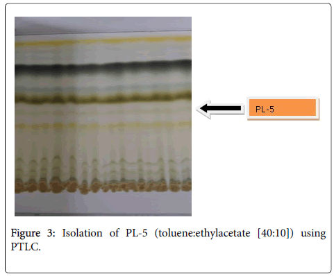
Figure 3: Isolation of PL-5 (toluene:ethylacetate [40:10]) using PTLC.
13C-NMR spectrum of compound PL-5 showed well resolved aliphatic, olefinic and carbonyl carbons of ester functional groups. 13CNMR of the compound revealed that the compound has 31 carbon atoms. By comparing the 13C-NMR and DEPT-135 NMR spectra there existed four quaternary carbon atoms with one oxygenated and one olefinic carbon at 173 and 142 ppm whereas the other quaternary carbons at δ 29.47 and 77.21 pm. The DEPT-135 NMR also displayed down ward peaks at δC, 24.49, 24.79, 25.03, 29.16, 29.28, 29.35, 29.69, 31.93, 34.42, 36.63, 37.29, 37.35, 37.42, 39.36, 39.85 and 61.19 ppm which showed the presence of sixteen methylene groups. In addition to that DEPT-135 NMR shows five methyl groups at 14.12, 16.36, 19.70, 19.74 and 22.62 ppm and six methine carbons at 22.68, 22.72, 27.97, 32.67, 32.80 and 118.15 ppm as upward peaks out of others. Based on the above information the compound PL-5 was proposed to have the following structure.
Antimicrobial assay
Antibacterial and antifungal activity test: The antimicrobial activities of crude extracts, isolated pure compounds and leaves oil were assayed by the presence or absence of inhibition zones diameter compared with some standard antibiotics in vitro against four bacteria (Table 3) and two fungi (Table 4). 10 μL and 20 μL of the crude extract and isolated compound (PL-5) were used for the study. The standard samples (chloramphenicol for bacteria and tilt for fungi) showed the greatest inhibition. The negative control solvent (petroleum ether) did not show any inhibition zone against tested fungi and bacteria (Figures 4 and 5).
| Sample | Dose (µl) | Types of bacteria with mean inhibition zone diameter (mm) | |||||
|---|---|---|---|---|---|---|---|
| Gram-negative bacteria | Gram positive bacteria | ||||||
| E. coli | S. thyphei | S. aureus | S. agalactiae | ||||
| 1 | 10 | 11 ± 0.10 | 5 ± 0.20 | 8 ± 0.10 | 11.2 ± 0.03 | ||
| 20 | 24 ± 0.30 | 24.5 ± 0.11 | 23 ± 0.20 | 17 ± 0.06 | |||
| 2 | 10 | 10 ± 0.01 | 2 ± 0.40 | 10 ± 0.16 | 11 ± 0.10 | ||
| 20 | 23 ± 0.10 | 24 ± 0.15 | 23 ± 0.3 | 19 ± 0.10 | |||
| 3 | 10 | 10 ± 0.08 | 3 ± 0.50 | 10 ± 0.10 | 9 ± 0.10 | ||
| 20 | 20.3 ± 0.10 | 24.5 ± 0.15 | 19.7 ± 0.30 | 16 ± 0.13 | |||
| 4 | 10 | 10 ± 0.61 | NI | 10 ± 0.24 | 7 ± 0.10 | ||
| 20 | 10 ± 0.12 | 11 ± 0.10 | 11 ± 0.30 | 9 ± 0.20 | |||
| 5 | 10 | 7 ± 0.40 | 2 ± 0.40 | 3 ± 0.50 | 7 ± 0.35 | ||
| 20 | 10 ± 0.20 | 9 ± 0.20 | 9 ± 0.10 | 9 ± 0.10 | |||
| 6 | 10 | NI | NI | NI | NI | ||
| 20 | NI | NI | NI | NI | |||
| 7 | 10 | 33.5 ± 0.03 | 25 ± 0.25 | 28 ± 0.09 | 31 ± 0.10 | ||
| 20 | 43 ± 0.09 | 43.5 ± 0.05 | 37.7 ± 0.14 | 32.2 ± 0.20 | |||
| Value represents mean of zone inhibition of three replication in mm ± SD; NI; Stands for No Inhibition |
|||||||
| Where 1 Pet. Ether crude extract 2 CHCl3/MeOH crude extract 3 MeOH crude extract 4 PL-5 |
5 Fr-6 6 Negative standards (Pet. Ether) 7 Positive standards (Chloramphenicol) |
||||||
Table 3: Zone of bacterial growth inhibition (mm) crude extracts, Fr-6 and PL-5 of Plantago lanceolata.
| Sample | Dose (µl) | Types of fungi with mean inhibition zone diameter (mm) | ||
|---|---|---|---|---|
| A. niger | F. solani | |||
| 1 | 10 | 18.2 ± 0.10 | 19.7 ± 0.23 | |
| 20 | 22 ± 0.10 | 20 ± 0.20 | ||
| 2 | 10 | 7.5 ± 0.09 | 8 ± 0.09 | |
| 20 | 10 ± 0.20 | 8 ± 0.10 | ||
| 3 | 10 | NI | 6.8 ± 0.08 | |
| 20 | NI | 8 ± 0.10 | ||
| 4 | 10 | NI | NI | |
| 20 | NI | NI | ||
| 5 | 10 | NI | NI | |
| 20 | NI | 8.2 ± 0.06 | ||
| 7 | 10 | NI | NI | |
| 20 | NI | NI | ||
| 8 | 10 | 32 ± 0.10 | 44 ± 0.40 | |
| 20 | 36 ± 0.11 | 50 ± 0.10 | ||
| Value represents mean of zone inhibition of three replication in mm ± SD; NI; Stands for No Inhibition | ||||
| Where 1 Pet. Ether crude extract 2 CHCl3/MeOH crude extract 3 MeOH crude extract 4 PL-5 |
5 Fr-6 6 Negative standards (Pet. Ether) 7 Positive standards (tilt) |
|||
Table 4: Zone of fungal growth inhibition (mm) crude extracts, Fr-6 and PL-5 of Plantago lanceolata.
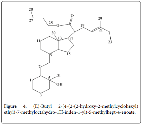
Figure 4: (E)-Butyl 2-(4-(2-(2-hydroxy-2-methylcyclohexyl) ethyl)-7-methyloctahydro-1H-inden-1-yl)-5-methylhept-4-enoate.
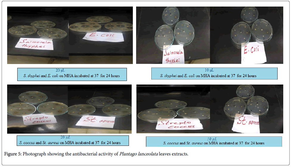
Figure 5: Photograph showing the antibacterial activity of Plantago lanceolata leaves extracts.
All crude extracts have higher inhibition effect against the tested bacteria in dose 20 μL when compared with dose 10 μL. PL-5 have lower inhibition effect against the tested bacteria in dose 20 μL compared with other all crude extract, which might be due to naturally occurring combinations of these components, might have synergistic effects [13]. Inhibition zones of the tested samples were to a little extent higher in gram negative bacterial (E. coli and S. thyphei ) than in gram positive bacterial (S. aureus and S. agalactiae ), hence, the tested samples have a stronger antibacterial activity towards gram negative than gram positive. The commercial standard drug (Chloramphenicol) showed the greatest inhibition effect against both tested bacteria in both doses (10 and 20 μL) compared with the tested samples and the negative control (solvent). The crude extracts and pure components are known to be active against a wide variety of microorganisms, including Gram-negative and Gram-positive bacteria. Therefore, leaves of Plantago lanceolata extracts are valuable and could be a future target for replacing synthetic antibacterial agents (Figure 6).
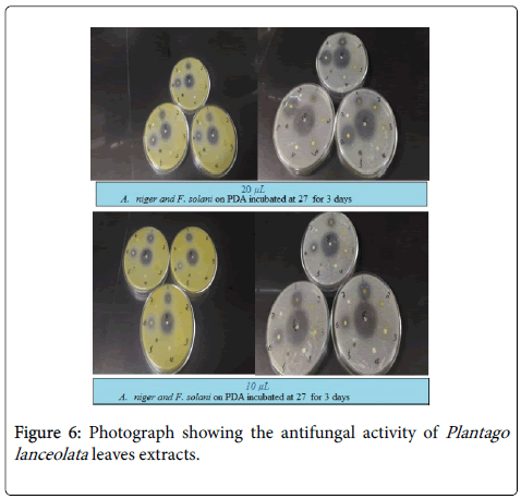
Figure 6: Photograph showing the antifungal activity of Plantago lanceolata leaves extracts.
The highest activity was determined in positive control (tilt) against F. solani 50 ± 0.10 mm the antifungal activities of other crude extracts were followed by petroleum ether, CHCl3/MeOH, MeOH and Fr-6 respectively. The lowest activity was determined in a MeOH against F. solani 6.8 ± 0.08 mm. In general the susceptibility of A. niger and F. solani to petroleum ether crude extract and CHCl3/MeOH crude extract demonstrate higher antifungal activities in terms of growth inhibition on the tested organisms make it a good candidate for investigation of in control of new multi-drug resistant on the tested fungi.
In this study the leaves P. lanceolata were extracted using petroleum ether, MeOH:CHCl3 (1:1) and MeOH. From the study, the leaves extract was found to contain steroids, alkaloids, flavonoids, tannins, saponins, glycosides, phenols and terpenoids. The presence of these important phytochemicals in the plant is a scientific justification of the plant use in the medicinal treatment against various diseases affecting humans. Additionally, crude extracts, isolated pure compound of the leaves were tested for the antimicrobial activities against four bacteria (Gram negative bacterium; E. coli and S. thyphei and the Grampositive bacterium; S. aureus and S. agalatiae ) and two fungi (A. niger and F. solani ). Antimicrobial activity tests showed some antimicrobial potency of the extracts when it was compared with standard antibiotics. After repeated successive solvent extraction and preparative thin layer chromatography different compounds were isolated from the plant leaf. Based on TLC analysis the plant contains several chemical constituents which were not isolated in this study because of financial and time constraints. The isolated pure compound coded as PL-5 was characterized using FT-IR, 1H-NMR, 13C-NMR and DEPT-135 analysis. Based on those spectral data, PL-5 was identified as one of non-aromatic steroid compound, (Tables 5-7) and Figures of Spectral data of compound PL-5.
| Observed frequency (cm-1) | Possible frequency range (cm-1) | Assignments |
|---|---|---|
| 3433 | 3200-3600 | -O-H stretching |
| 2922 | 2850-3000 | Asymmetric -C-H3 stretching of alkanes |
| 2853 | 2850-2950 | Symmetric -C-H2 stretching of alkanes |
| 1754 | 1665-1760 | Carboxyl compound |
| 1464 | 1350-1470 | -C-H2 bending for alkanes |
| 693 | 665-1000 | -C-H bending (unsubstituted alkene) |
Table 5: Typical Infra-Red (IR) absorption frequencies of the compound PL-5.
| Chemical shift of PL-5 |
Multiplicity, No of Hydrogen |
Literature value | Remark |
|---|---|---|---|
| 0.78-0.96 | Multiplet, 15H | 0.80-1.28 | CH3- |
| 1.02-1.208 | Multiplet, 18H | 1.00-1.44 | Cycloalkanes proton |
| 1.50-1.58 | Multiplet, 12H | 1.50 | -CH2- |
| 1.60-1.96 | Singlet, 4H | 1.40-1.80 | -CH- |
| 2.27-2.28 | Triplet, 1H (J=15.2 Hz) | 0.50-2.00 | -CH- |
| 2.77-2.80 | Singlet, 1H | 2.25-3.50 | Hydroxyl proton |
| 4.43-4.58 | Doublet, 1H (J=7.2 Hz) | 4.20-5.00 | -OCH2- |
| 5.02-5.314 | Multiplet, 1H | 5.00-5.3 | Olefinic proton |
Table 6: 1H-NMR spectral data of PL-5 in CDCl3.
| Carbon position | 13C-NMR d (ppm) | DEPT-135 d (ppm) | Remark |
|---|---|---|---|
| Me-23 | 14.12 | 14.12 | -CH3 |
| Me-30 | 16.36 | 16.37 | -CH3 |
| Me-28 | 19.70 | 19.71 | -CH3 |
| Me-29 | 19.74 | 19.75 | -CH3 |
| Me-31 | 22.62 | 22.63 | -CH3 |
| C-9 | 22.68 | 22.69 | -CH- |
| C-14 | 22.72 | 22.72 | -CH- |
| C-27 | 24.49 | 24.47 | -CH2 |
| C-11 | 24.79 | 24.80 | -CH2 |
| C-3 | 25.03 | 25.03 | -CH2 |
| C-6 | 27.97 | 27.98 | -CH- |
| C-15 | 29.16 | 29.15 | -CH2 |
| C-19 | 29.28 | 29.27 | -CH2 |
| C-2 | 29.35 | 29.35 | -CH2 |
| C-13 | 29.47 | - | Quaternary |
| C-8 | 29.69 | 29.65 | -CH2 |
| C-1 | 31.93 | 31.92 | -CH2 |
| C-18 | 32.67 | 32.66 | -CH- |
| C-17 | 32.80 | 32.79 | -CH- |
| C-16 | 34.42 | 34.40 | -CH2 |
| C-22 | 36.63 | 36.63 | -CH2 |
| C-26 | 37.29 | 37.30 | -CH2 |
| C-10 | 37.35 | 37.36 | -CH2 |
| C-7 | 37.42 | 37.44 | -CH2 |
| C-12 | 39.36 | 39.37 | -CH2 |
| C-4 | 39.85 | 39.86 | -CH2 |
| C-25 | 61.19 | 61.20 | -OCH2 |
| C-5 | 77.21 | - | Quaternary |
| C-20 | 118.15 | 118.16 | -CH- |
| C-21 | 142.60 | - | Quaternary |
| C-24 | 173.94 | - | Quaternary |
Table 7: 13C-NMR and DEPT-135 spectral data of PL-5 in CDCl3.
My sincere gratitude is given to Ethiopian Ministry of Education for financial support. I am also very much grateful to my major advisor Neelaiah Babu G (PhD) for his support on laboratory works. My thanks also go to for all who supported me by sharing idea or offering the material which is an input for this work.
