Research Article - (2015) Volume 3, Issue 2
Aframomum melegueta is an herb in the ginger family that has been shown to have anti-inflammatory, antioxidative, anti-diabetic and antimicrobial properties. We investigated the possibility that the seeds of this herb, which are consumed by gorillas and used as a spice in West and North African cuisine, could have neuroprotective effects in a rat model of traumatic brain injury (TBI). Using Fluoro-Jade, an anionic fluorescent stain that is a well-established marker of degenerating neurons, we found that an extract of Aframomum, PMI-006, significantly reduced numbers of dying, Fluoro-Jade-positive neurons in the rat hippocampus 24 hr after TBI. We used an antibody to CD11b (Ox42), a microglial marker, to show that PMI almost completely reduced microglial activation-a hallmark of injury-induced inflammation- in the rat hippocampus and cortex. To elucidate the molecular mechanisms underlying the neuro protective effects of PMI-006, we used RT2 Profiler pathway-focused PCR arrays representing oxidative stress, cytokine & chemokines and NFκB cell signaling pathways to interrogate PMI-induced changes in hippocampal gene expression after TBI. We found that PMI treatment ameliorated the effects of brain injury and, in several cases, restored injuryinduced gene expression changes to sham control levels. PMI treatment did not significantly alter functional outcome in the Morris Water Maze, a neurobehavioral test of hippocampal-dependent spatial memory. However, because of its safety profile and because it mitigates the effects of TBI on stress and inflammatory signaling pathways that are associated with TBI pathology, PMI could be potentially beneficial in reducing neurodegeneration in TBI survivors.
Keywords: Traumatic brain injury; Aframomum melegueta; Hippocampus; Inflammatory genes; Oxidative stress genes; NFkB signaling genes; Fluoro-Jade
Natural products and their derivatives were the basis for early medicines and continue to provide a rich source of drugs today, with up to 60% of approved new chemical entities (NCE) originating from natural sources [1–5]. A major reason for this is thought to be that the small molecules produced by diverse organisms have evolved to interact with biological targets which are broadly conserved across the animal and plant kingdoms, and that these compounds are generated and maintained in diverse lineages of living organisms [4].
Aframomummelegueta is a species in the ginger family, Zingiberaceae, members of which are known to possess strong antiinflammatory and/or antibacterial properties [6,7]. In West African folk medicine, Aframomummelegueta seeds, known as Grains of Paradise, are valued for their warming and digestive properties as well as numerous medicinal effects. Researchers in a biotechnology company, Phytomedics, found that a derivative of this plant, PMI-006, has powerful anti-inflammatory effects, comparable to the well-known anti-inflammatory drugs Vioxx, Celebrex and Bextra but without their adverse side effects.Thus, it has been suggested that Aframomum might successfully be used to treat diseases with inflammation as their hallmarks, such as cardiovascular conditions, arthritis, osteoporosis and Alzheimer's disease.Anecdotal evidence also suggests that for centuries, native African healers have used Aframomumto treat infections of all kinds. Recent studies have provided experimental support for these antimicrobial effects [8]. Because of these medicinal properties, we reasoned that this compound may prove neuro protective in our rat model of fluid percussion traumatic brain injury (TBI), which is a clinically relevant model for human brain injury [9-11].
TBI is a leading cause of death and lifelong disability, but there are no approved drugs for millions of TBI survivors due to the failure of all pharmacotherapeutic treatments in clinical trials [12–14]. Thus, it is imperative that we continue to explore novel sources of drug candidates for TBI. In millions of patients surviving civilian and military trauma, the costs of TBI include disruption of daily functions, irreparable cognitive impairment, inability to return to work and overall decreased quality of life [15]. Thornhill et al. [16] evaluated 459 survivors of mild, moderate and severe TBI at one year after injury and reported that even the mildly injured patients had a 43% incidence of cognitive impairment, much of which is associated with injury-induced neurodegeneration in the hippocampus, a region in the medial temporal lobe that is critical to learning, memory and executive function [17,18]. The critical role of the hippocampus in brain function is evident in neurological disorders that are associated with cognitive dysfunction. Memory loss and dementia in Alzheimer’s patients are closely correlated with loss of hippocampal neurons, and TBI patients commonly experience memory and learning deficits that are linked to hippocampal damage [19-21]. Functional neuroimaging studies have shown that the hippocampus is actively engaged during navigational tasks in humans and that hippocampal damage directly influences its interactions with other brain regions during memory retrieval [22,23].
The purpose of this study was to determine if an extract derived from Aframomummelegueta(PMI-006) could improve functional outcome after experimental TBI and if so, to identify underlying mechanisms of neuro protection. Using an established fluorescent marker of degenerating neurons, Fluoro-Jade [24], immune histochemical analysis of activated microglia, gene expression analysis using pathwayfocused PCR arrays and a test of hippocampal-dependent cognitive function, the Morris Water Maze [25], we characterized the neuro protective effects and therapeutic potential of this natural productderived compound.
Preparation of Aframomummelegueta
PMI-006 extract was obtained from Phytomedics, Inc (Jamesburg, NJ). Preparation of the extract has been described by Ilic et al. [26]. Phytomedics scientists have used PMI 006 at doses 250-1000 mg/kg and saw physiological effects during the next 24 hours. From toxicology tests, they determined that NOAEL (no-observed-adverse-effect-level) is 1500 mg/kg. Experimental results generated at Phytomedics showed that it takes 3 hours of pretreatment for PMI-006 to be effective. All of PMI botanical drugs are administered orally. Per Phytomedics suggestions, we solubilized PMI-006 in 100% ethanol and prepared a 10% ethanol solution brought to volume with corn oil and administered it to our experimental rats via oral gavage 1 hour after injury.
Fluid percussion injury
The Institutional Animal Care and Use Committee of The University of Texas Medical Branch approved all experimental protocols. Adult, male, Charles River Sprague-Dawley rats (300-400g) were anesthetized with 4% isoflurane, intubated, then mechanically ventilated and prepared for fluid percussion TBI.A craniotomy was performed laterally to the sagittal suture, midway between the lambda and bregma structures. The fluid percussion device was then attached, and the animal was subjected to severe lateral fluid percussion traumatic brain injury (TBI) as previously described [10]. The rats were sacrificed 24hrs post-injury for neuronal counting of degenerating neurons [27] and PCR array analysis. For immunohistochemistry, rat brains were dissected out eleven days after injury following the final day of Morris Water Maze testing, immediately frozen on dry ice, and stored at -80°C.
Neuronal counting
Rats were randomly assigned to receive TBI plus 10mg, 100mg, 250mg, 500mg or 1000mg of PMI-006 (mg/kg body weight) or TBI plus vehicle alone (10% ethanol in corn oil) via oral gavage. Animals were survived for 24
Hours postin jury, sacrificed, their brains removed, sectioned on acryostatand10 μM Frozen coronal brain sections were stained with Fluoro–Jade [27] and counterstainedwithanisslstain,1% cresyl violet. A blinded investigator then counted Fluoro-Jade positive neurons in the CA1/2 and CA3 regions on the ipsilateral (injured) side of the rat hippocampus imaged using an Olympus BX51 Fluorescent Microscope. The numbers of FJ-positive neurons were quantified for each of the treatment groups and reported as mean +/− SEM and analyzed using an analysis of variance (ANOVA) followed by the Bonferroni-Dunn test with α=0.05. Statistical computations were carried out using PROC GLM in SAS®, Release9.1[28].
Neurobehavioral assessment using Morris Water Maze (MWM)
MWM procedures assessing working memory are described in detail by Hamm et al. [25]. Tank parameters for the MWM were as follows: a black tank (180 cm diameter, 28 cm depth) was filled with ambient temperature water. When filled, the tank contained a clear plastic platform hidden beneath the surface of the water. Acquisition blocks consisted of two daily trials over five consecutive days (7-11 days after injury). At the onset of each acquisition trial, rats were placed by hand in the pool facing the tank wall. There were four zones and four starting areas for the rats; each rat was given two trials at each starting point. In this version of the Morris water maze, which assessed working memory, the location zone of the hidden platform was randomized between trials on the same day.Animals (10 sham, 8 TBI, and 12 TBI + PMI-006) were allowed to swim a maximum of 2 min to find the hidden platform and the latency was recorded for each trial. If the rat failed to find the platform after 2 min, it was placed on the platform by the experimenter. All rats were allowed to remain on the platform for 15 sec before being returned to a heated holding box for a 4 min intertrial interval. The SMART computer program (SMART program, San Diego Instruments, Inc., San Diego, CA) was used to collect, store and analyze the behavioral data. Statistical analyses were performed using PROC MIXED in SAS® (version 9.4) with no adjustment for covariance necessary and a Tukey adjustment for multiple comparisons.
Immunohistochemistry for assessment of microglial activation
To assess the effects of PMI-006 on TBI-induced inflammation, we performed immune histochemical analysis of TBI, PMI-006 treated and sham control rat brains using an antibody to CD11b (OX-42), a marker of microglial activation. Eleven days after injury, rats were sacrificed (n=3/group), perfused with 4% paraformaldehyde, brains collected and 10 μm frozen sections were cut on a cryostat. Sections were then incubated overnight with a 1° antibody (mouse anti-CD11b; 1:2000, BD Biosciences, San Jose, CA). The following morning, sections were incubated with a 2°antibody (Alexa 594 goat anti-mouse; 1:400, Life Technologies, Grand Island, NY) at ambient temperature, and then mounted with DAPI (stains nuclei) for imaging. An Olympus BX51 Fluorescent Microscope was used to visualize the hippocampal formation and surrounding cortical regions.
RNA isolation
Total RNA was isolated from dissected ipsilateral (injured side) hippocampal tissue samples using the Ultraspec RNA isolation System (Biotecx Laboratories, Inc. Houston, TX) following the manufacturer’s protocol for RNA isolation from whole tissue. Genomic DNA contamination was removed by with DNase treatment (Ambion, Austin TX) and then RNA was ethanol precipitated and brought up in nuclease-free water.RNA from TBI, TBI + PMI-006 treated, and sham injured animals was assessed for quality and quantity on an Agilent Bioanalyzer (Agilent Technologies, Santa Clara CA) with RIN values consistently averaging 7.0 to 8.0 or higher.Approximately 1 μg of each RNA sample was reverse transcribed using the RT2 First Strand Kit (SA Biosciences) in preparation for use in PCR arrays.
RTProfiler PCR arrays
To profile the expression of genes related to oxidative stress, cytokines and chemokines, and NF-κB signaling pathways after TBI, TBI+PMI-006 and sham injury, quantitative real- time PCR was performed using the RT2 Sybr-green Profiler PCR Arrays (SA Biosciences, Valencia, CA) following manufacturer’s protocols. The expression of genes involved in the oxidative stress, cytokine & chemokine, and NF-κB signaling pathways (n=3, 4, 3/per group, respectively) in the TBI alone and TBI + PMI-006 treated rat brains were compared to the baseline levels of the same genes in the sham injured rat brains. Data analysis was based on the delta-delta CT method with normalization of the raw data to 5 housekeeping genes and calculations of the fold changes were done using the Analysis Web portal program provided by SA Biosciences.
PMI-006 reduces neuronal injury in rat hippocampus
To determine the lowest dose of PMI-006 that provides maximal protective effects, we performed a dose response. In animalstreatedwithPMI-006, there was a significant decrease in the number of Fluoro-Jade positive neurons in groups (250, 500, 1000 mg/kg) compared with vehicle treatment alone in the CA1/2 region suggesting that PMI 006 reduces neuronal injury in the hippocampus (Figure 1A, B). For Figure 1A and all subsequent experiments, we used a dose of 250mg/kg, the lowest dose which was associated with the greatest reduction in neuronal injury.
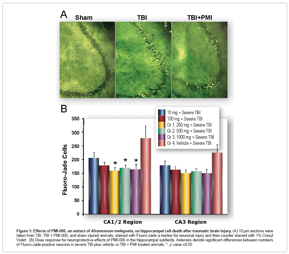
Figure 1: Effects of PMI-006, an extract of Aframomum melegueta, on hippocampal cell death after traumatic brain injury. (A) 10 μm sections were taken from TBI, TBI + PMI-006, and sham injured animals, stained with Fluoro-Jade a marker for neuronal injury and then counter stained with 1% Cresyl Violet. (B) Dose response for neuroprotective effects of PMI-006 in the hippocampal subfields. Asterisks denote significant differences between numbers of Fluoro-Jade-positive neurons in severe TBI plus vehicle vs TBI + PMI treated animals; *: p value ≤0.05
PMI-006 reduces microglial activation
Because microglial activation is a hallmark of TBI [29], we performed immune histo chemical analysis of injured rat brain sections using an antibody to CD11b, a marker of activated microglia. PMI-006 had a remarkably ameliorative effect on microglial activation after TBI (Figure 2), almost completely abolishing injury-induced inflammation in the hippocampus and cortex.
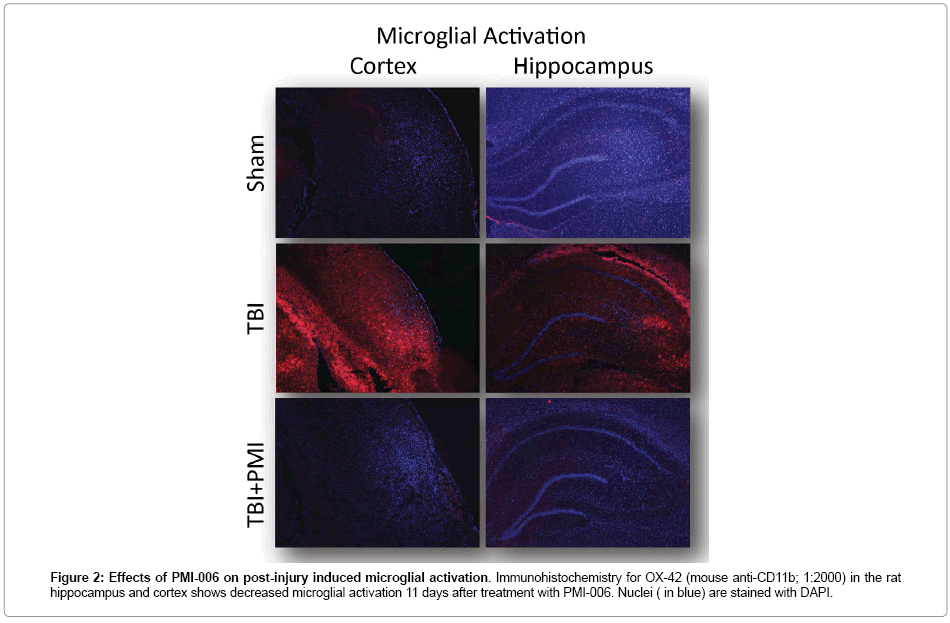
Figure 2: Effects of PMI-006 on post-injury induced microglial activation. Immunohistochemistry for OX-42 (mouse anti-CD11b; 1:2000) in the rat hippocampus and cortex shows decreased microglial activation 11 days after treatment with PMI-006. Nuclei ( in blue) are stained with DAPI.
Pathway-focused PCR array analysis
The PCR super arrays (Oxidative stress, Cytokine and Chemokines, and NFκB pathways) were chosen because of the reported antiinflammatory and anti-oxidative properties of PMI-006. In all three PCR array data sets, we found that, as expected, severe TBI alone induced multiple proinflammatory and oxidative stress-inducing genes (complete dataset of fold changes are shown in Tables S1, S2 and S3). In each pathwayfocused array, we compared significant changes induced by PMI treatment to significant changes induced by TBI alone (Figure 3). Most of these significantly affected genes are associated with injury-associated pathways that are implicated in the pathogenesis of TBI (Supplemental References).In most of these cases, we observed a consistent and reproducible trend: PMI-006 treatment reversed or normalized the effects of TBI in the direction of sham control levels.Interestingly, these gene expression resultssuggested that PMI-006 mitigated the effects of TBI on both deleterious genes and some protective genes, such as GPx-1, that are involved in the brain’s endogenous protectiveresponses toinjury [30].
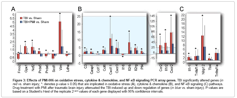
Figure 3: Effects of PMI-006 on oxidative stress, cytokine & chemokine, and NF-κB signaling PCR array genes. TBI significantly altered genes (in red vs. sham injury; *: denotes p-value ≤ 0.05) that are implicated in oxidative stress (A), cytokine & chemokine (B), and NF-κB signaling (C) pathways. Drug treatment with PMI after traumatic brain injury attenuated the TBI-induced up and down regulation of genes (in blue vs. sham injury). P-values are based on a Student’s t-test of the replicate 2(-ΔCt) values of each gene displayed with 95% confidence intervals.
Morris water maze
We examined the effect of PMI-006 on neuro behavioral outcome after TBI using the Morris Water Maze, an
Established test of hippo campal-dependent working memory (Figure 4). There were no apparent significant differences between any of the comparisons. Althoughsignificant differences were not apparent, there was a borderline significant difference between Sham and TBI (p=0.0526).The other comparisons (Sham vs. PMI, p=0.2846, PMI larger; TBI vs. PMI, p=0.7338 with TBI larger) were not significant. There were no interactions of note (interaction of day and group: p=0.3801).As for days, day 7 was largest (p<0.001 comparing to all others); consecutive days were not significantly different (8 vs. 9, p=0.5324; 9 vs. 10, p=0.4935; 10 vs. 11, p=0.2572), but nonconsecutive days were significantly different (8 vs. 10, p=0.0142; 9 vs. 11 p=0.0028). The effect was decreasing over time. There was also a significant effect of trial (p<0.0001).
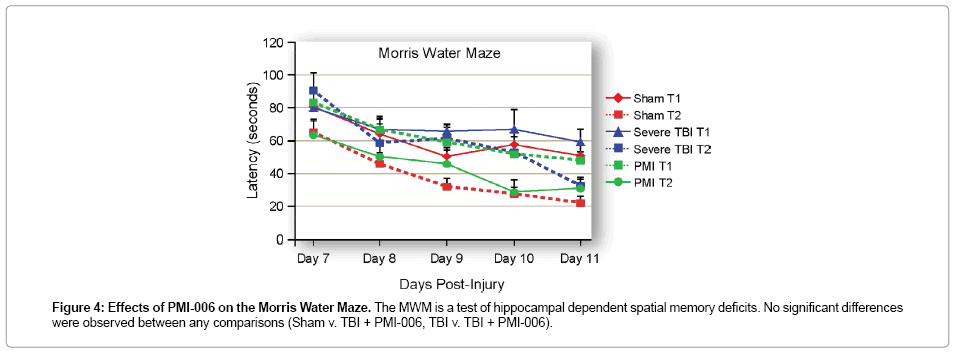
Figure 4: Effects of PMI-006 on the Morris Water Maze. The MWM is a test of hippocampal dependent spatial memory deficits. No significant differences were observed between any comparisons (Sham v. TBI + PMI-006, TBI v. TBI + PMI-006).
Despite decades of brain injury research and demonstrated success in pre-clinical studies using animal models of
TBI, numerous clinical trials of neuro protective agents have failed to show efficacy in human TBI patients [13,31].Thus, we and others are still searching for novel therapeutic treatments that have the potential to mitigate TBI-inducedneurodegenerationandimprovefunctionaloutc omeinTBIsurvivors. It is well established that injury-
Induced inflammation and oxidative stress contribute significantly to deleterious secondary injury signaling cascades that result in progressive long-term neuro degeneration that is associated with lifelong disability in human TBI survivors [32,33].Therapeutic treatments that affect these pathways could reduce neuronal death and improve functional outcome.
Here, we have described the molecular and functional effects of a new therapeutic drug candidate, PMI-006, a natural compound derived from the seeds of the Aframomummelegueta plant. Several studies have shown that extracts of Aframomummelegueta have strong anti-oxidant [34,35], anti-microbial [7,8], anti-apoptotic [6], anti-diabetic [36], antinociceptive [37] and anti-inflammatory [38] properties. Cumulatively, these properties suggest that this compound could have both analgesic and neuro protective effects after TBI. Our data is entirely consistent with these previous observations. The reduction of neuronal injury in the hippocampus correlates with the ameliorative effects of PMI treatment on TBI-induced inflammatory and oxidative stress signaling. Evidence suggests that drugs with pleiotropic properties, i.e., that possess both anti-inflammatory and anti-oxidative effects, appear to significantly improve functional outcome after TBI [39].Because neuronal death from brain injury is due, in part, to a strong inflammatory response in the damaged brain tissue, these data support our hypothesis that natural product derived compounds with potent anti-inflammatory properties such as PMI 006 may reduce neuronal injury.
The demonstrated safety profile of Aframomummelegueta (e.g., its consumption by animals and humans) as well as its demonstrated protective effects in experimental models of human disease suggests that this compound is an excellent candidate for translational TBI studies. The potent neuro protective properties of PMI may be due, in part, to its anti-inflammatory effects but PCR array data also show that neuro protection may be the result of its alteration of pro- and anti-oxidant pathways and its effects on NFκB signaling (which is known to be activated after head injury) [40]. Although these studies were conducted in a rodent model of TBI, it has been shown that gene expression changes after TBI are commonly modulated across different species [41], suggesting that similar effects would be expected in human TBI patients. Natural compounds, such as PMI-006, that demonstrate such properties have great therapeutic potential for reducing neurodegeneration and improving functional outcome in TBI patients.
This study was supported by R21 NS053620, RO1 NS053620, the Moody foundation, the University of Texas Medical Branch at Galveston Department of Anesthesiology, and the Medical student research program. We thank Christine Courteau-Butler and Andy Hall for excellent editorial support and Christy Perry for figures and illustrations. We thank Phytomedics, Inc. for freely and generously providing the Aframomummelegueta extract for our experimental brain injury studies.
